Wed, May 27, 2026
Volume 14, Issue 2 (Summer & Autumn 2017)
ASJ 2017, 14(2): 79-82 |
Back to browse issues page

Download citation:
BibTeX | RIS | EndNote | Medlars | ProCite | Reference Manager | RefWorks
Send citation to:


BibTeX | RIS | EndNote | Medlars | ProCite | Reference Manager | RefWorks
Send citation to:
Jalali Kondori B, Asadi M H, Bahadoran H, Dadseresht S. Anthropometric Study of Hip Joint in Tehran Population Using Computed Tomography Scan. ASJ 2017; 14 (2) :79-82
URL: http://anatomyjournal.ir/article-1-191-en.html
URL: http://anatomyjournal.ir/article-1-191-en.html


1- Department of Anatomical Sciences, School of Medical Sciences, Baqiyatallah University of Medical Sciences, Tehran, Iran.
Full-Text [PDF 428 kb]
(1944 Downloads)
| Abstract (HTML) (5805 Views)


Full-Text: (2655 Views)
1. Introduction
Studies indicate that anatomic parameters of hip joint differ among various populations. Detailed study of hip joint using anthropometric studies could help us specify its biomechanical functions. In addition, the early pathological changes of joint in some diseases such as primary osteoarthritis can be diagnosed by determining normal values of anatomical components of hip joint in each population [1, 2]. The Computed Tomography (CT) is a valuable technique in studying human anatomy. In this method besides viewing the entire structure of hip joint, some quantitative information is obtained about the size, angle, thickness, and other parameters of joint structures [3]. Determining the normal parameters and angles in a population results in better identification of abnormal values and finding their relationships with the incidence and prevalence of skeletal diseases in the population.
This study aimed to evaluate the anatomical variations in the normal hip joint and determine its anthropometric parameters using CT scan in Tehran population and then comparing these values with the same parameters in other populations.
2. Materials and Methods
This study was conducted on the hip joints of 260 patients in all ages and both genders who had a pelvic CT scan in the Department of Radiology at Ebn-e Sina Hospital, Tehran, Iran between April 2014 to September 2015. The average age of the patients was 42 years and their age range was between 25 and 65 years. Those patients with the history of operations on pelvic and lower extremity and those with the center edge angle of less than 20 degrees were excluded from the study.
In this study, we assessed four parameters in hip joint using CT scan technique. In all cases, the patients lied in the supine position and their lower limbs were in fully extended position. The patients’ legs were fixed by a strap. First, a topogram was taken from the top of hip joint to the bottom of the femoral shaft. The axial cuts with a thickness of 5 mm were prepared from the hip joint area.
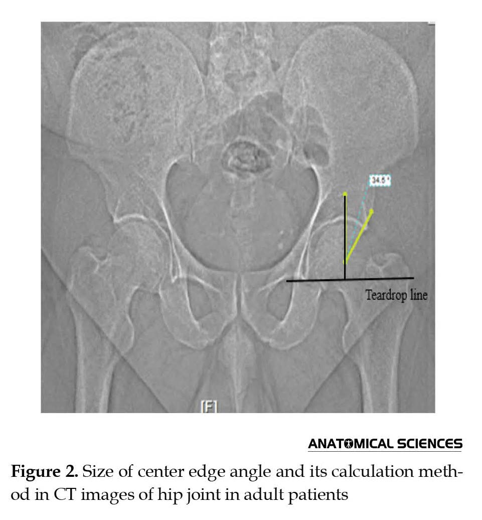
The parameters used in this study are defined as follows: 1. Center Edge (CE) angle of Wiberg which is the angle formed between the perpendicular line passing through the center of the femoral head and the line drawn through the center of the femoral head to the anterior acetabular edge; 2. Neck shaft angle which is the angle formed between the longitudinal axis of the femoral neck and the longitudinal axis of the femoral shaft; 3. Acetabular angle of sharp which is the angle formed between the horizontal line passing from the tip of the pelvic teardrop and the line drawn through the tip of teardrop to anterior acetabular edge; 4. Acetabular depth which is distance between the center of line drawn from the anterior acetabular margin to lower acetabular margin and acetabular apex (Figure 1).
Studies indicate that anatomic parameters of hip joint differ among various populations. Detailed study of hip joint using anthropometric studies could help us specify its biomechanical functions. In addition, the early pathological changes of joint in some diseases such as primary osteoarthritis can be diagnosed by determining normal values of anatomical components of hip joint in each population [1, 2]. The Computed Tomography (CT) is a valuable technique in studying human anatomy. In this method besides viewing the entire structure of hip joint, some quantitative information is obtained about the size, angle, thickness, and other parameters of joint structures [3]. Determining the normal parameters and angles in a population results in better identification of abnormal values and finding their relationships with the incidence and prevalence of skeletal diseases in the population.
This study aimed to evaluate the anatomical variations in the normal hip joint and determine its anthropometric parameters using CT scan in Tehran population and then comparing these values with the same parameters in other populations.
2. Materials and Methods
This study was conducted on the hip joints of 260 patients in all ages and both genders who had a pelvic CT scan in the Department of Radiology at Ebn-e Sina Hospital, Tehran, Iran between April 2014 to September 2015. The average age of the patients was 42 years and their age range was between 25 and 65 years. Those patients with the history of operations on pelvic and lower extremity and those with the center edge angle of less than 20 degrees were excluded from the study.
In this study, we assessed four parameters in hip joint using CT scan technique. In all cases, the patients lied in the supine position and their lower limbs were in fully extended position. The patients’ legs were fixed by a strap. First, a topogram was taken from the top of hip joint to the bottom of the femoral shaft. The axial cuts with a thickness of 5 mm were prepared from the hip joint area.
The parameters used in this study are defined as follows: 1. Center Edge (CE) angle of Wiberg which is the angle formed between the perpendicular line passing through the center of the femoral head and the line drawn through the center of the femoral head to the anterior acetabular edge; 2. Neck shaft angle which is the angle formed between the longitudinal axis of the femoral neck and the longitudinal axis of the femoral shaft; 3. Acetabular angle of sharp which is the angle formed between the horizontal line passing from the tip of the pelvic teardrop and the line drawn through the tip of teardrop to anterior acetabular edge; 4. Acetabular depth which is distance between the center of line drawn from the anterior acetabular margin to lower acetabular margin and acetabular apex (Figure 1).
The obtained data were analyzed by SPSS and the different results were investigated in each gender. In our study, P value less than 0.05 was considered as significant.
3. Results
The mean obtained center edge angle was 32 degrees. The range of this angle was 27-47 degrees in the studied patients (Figure 2). The angle between the longitudinal axes of the femoral neck and shaft was also measured in all patients. The mean neck shaft angle was 139.5 degrees. This angle ranged from 137 to 146 degrees. An example of how to calculate the angle in the CT scan images is given in Figure 3. In this study, the mean acetabular angle was 37.1 degrees and ranged from 32 to 41 degrees (Figure 4). In our research, the mean depth of acetabular was 1.67 cm. This parameter ranged between 1.93 to 1.52 cm (Figure 5).
Assessment of the differences among parameters between genders showed that the neck shaft angle in women was about 2.5 degrees more than that in men. Other parameters showed no significant differences between men and women.
4. Discussion
Anthropometric studies can provide valuable information on different parameters of bones, joints, and their variations in different populations. Assessment of anatomic status, bone shape, and bone density can be performed much better using CT scan technique. With regard to 3D simulation ability of multi-slice CT scanning device, its application seems very useful in anthropometric studies. In this study, we measured the different parameters of hip joint in adults living in Tehran, Iran using CT scan technique, so that we could quantitatively compare these parameters in other populations. In addition, the quantitative comparison was also performed between genders. The normal CE angle is more than 20 degrees and lower than this is considered as dysplasia [4]. In a similar study conducted by Shi on Chinese people, the mean CE angle was reported as 32.8 degrees with a range between 13.7 and 58.8 degrees [5]. In Saikia et al. study, the average CE angle was 32.7 degrees with a range of 60-20 degrees [6]. The results of these two studies regarding CE angle are in line with our findings.
Previous studies have numerous reports on neck shaft angle [7, 8]. For example, in Lequesne study, the mean angle has been reported as 132.8 degrees [9]. Also in Hoaglund research, this angle has been reported as 135 degrees in adults [10]. In our study, the angle was 139.5 degrees in adults that is slightly larger than other populations. According to our results, the size of this angle in women is 2.5 degrees more than that in men.
Acetabular angle was reported by Sharp for the first time. This angle is calculated to determine dysplastic joints. Values greater than 43 degrees are considered as dysplasia. In a study conducted by Nakamura on Japanese people, this angle has been reported as 38 degrees [11]. Also in Saikia study, this angle was reported as 39.2 degrees [6]. This angle in our study population is similar to these studies results with no significant difference.
Acetabular depth is one of the most important parameters to determine acetabular dysplasia, i.e., values less than 9 mm are considered as acetabular dysplasia [12]. In our study, this depth ranged between 1.93 and 1.52 cm and there was no case of acetabular dysplasia. Knowledge on the anthropometric parameters of main bones and joints such as hip joint could remarkably help in diagnosis of diseases related to the skeletal system [13].
The results of the present study performed using computed topography techniques suggest that the majority of anthropometric parameters regarding hip joint in the study population are similar to other populations, though the neck shaft angle in this study was a few degrees larger than that in other populations. Moreover, the mentioned angle in females was 2.5 degrees larger than that in males.
Acknowledgments
This research did not receive any specific grant from funding agencies in the public, commercial, or not-for profit sectors. We would like to thank Dr. Mohamad Hossein Asadi, head of Department of Anatomical Sciences, Baqiyatallah University of Medical Sciences, for his guidance and support in this study.
Conflict of Interest
The authors declared no conflicts of interest.
References
- Lequesne MG, Laredo JD. The faux profil (oblique view) of the hip in the standing position: Contribution to the evaluation of osteoarthritis of the adult hip. Annals of the Rheumatic Diseases. 1998; 57(11):676–81. doi: 10.1136/ard.57.11.676
- Chauhan R, Paul S, Dhaon BK. Anatomical parameters of North Indian hip joints: Cadaveric study. Journal of the Anatomical Society of India. 2002; 51(1):39-42.
- Feldkamp LA, Goldstein SA, Parfitt MA, Jesion G, Kleerekoper M. The direct examination of three-dimensional bone architecture in vitro by computed tomography. Journal of Bone and Mineral Research. 2009; 4(1):3–11. doi: 10.1002/jbmr.5650040103
- Wiberg G. Studies on dysplastic acetabula and congenital subluxation of the hip joint: With special reference to the complication of osteoarthritis. Acta Chirurgica Scandinavica. 1939; 83(58):53-68.
- Shi YY, Liu TJ, Zhao Q, Zhang LJ, Ji SJ, Wang EB. The normal centre-edge angle of Wiberg in the Chinese population: A population-based cross-sectional study. Journal of Bone and Joint Surgery - British Volume. 2010; 92-B(8):1144–7. doi: 10.1302/0301-620x.92b8.23993
- Saikia K, Bhuyan S, Rongphar R. Anthropometric study of the hip joint in Northeastern region population with computed tomography scan. Indian Journal of Orthopaedics. 2008; 42(3):260-6. doi: 10.4103/0019-5413.39572
- Standring S. Gray's anatomy: The anatomical basis of clinical practice. New York: Churchill Livingstone; 2000.
- Adekoya Cole T, Akinmokun O, Soyebi K, Oguche O. Femoral neck shaft angles: A radiological anthropometry study. Nigerian Postgraduate Medical Journal. 2016; 23(1):17. doi: 10.4103/1117-1936.180130
- Lequesne MG, Laredo JD. The faux profil (oblique view) of the hip in the standing position. Contribution to the evaluation of osteoarthritis of the adult hip. Annals of the Rheumatic Diseases. 1998; 57(11):676–81. doi: 10.1136/ard.57.11.676
- Hoaglund FT, Djin W. Anatomy of the femoral neck and head, with comparative data from Caucasians and Hong Kong Chinese. Clinical Orthopaedics and Related Research. 1980; 152:10-6. doi: 10.1097/00003086-198010000-00003
- Nakamura S, Ninomiya S, Nakamura T. Primary osteoarthritis of the hip joint in Japan. Clinical Orthopaedics and Related Research. 1989; 241:190-6. doi: 10.1097/00003086-198904000-00021
- Loder RT, Mehbod AA, Meyer C, Meisterling M. Acetabular depth and race in young adults. Journal of Pediatric Orthopaedics. 2003; 23(6):699–702. doi: 10.1097/01241398-200311000-00003
- Jain A, Maheshwari A, Singh M, Nath S, Bhargava S. Femoral neck anteversion: A comprehensive Indian study. Indian Journal of Orthopaedics. 2005; 39(3):137-44. doi: 10.4103/0019-5413.36685
Type of Study: Original |
Subject:
Morphometry
Received: 2016/09/5 | Accepted: 2017/03/18 | Published: 2017/07/1
Received: 2016/09/5 | Accepted: 2017/03/18 | Published: 2017/07/1
Send email to the article author
| Rights and permissions | |
 |
This work is licensed under a Creative Commons Attribution-NonCommercial 4.0 International License. |

Copyright © The Author(s);
This is an open access article distributed under the terms of the Creative Commons Attribution License (CC-By-NC), which permits use, distribution, and reproduction in any medium, provided the original work is properly cited and is not used for commercial purposes.
Contact Information
