Sat, Jul 25, 2026
Volume 20, Issue 2 (Summer & Autumn 2023)
ASJ 2023, 20(2): 67-78 |
Back to browse issues page


![]()
![]()
![]()
Download citation:
BibTeX | RIS | EndNote | Medlars | ProCite | Reference Manager | RefWorks
Send citation to:


BibTeX | RIS | EndNote | Medlars | ProCite | Reference Manager | RefWorks
Send citation to:
Gharaei P, Safi S, Hesaraki S, Kakoienejad M M. Biochemical and Histopathological Studies on the Protective Effects of Aged Garlic Extract on Heart and Kidney Injuries Caused by Doxorubicin in Female Mice. ASJ 2023; 20 (2) :67-78
URL: http://anatomyjournal.ir/article-1-629-en.html
URL: http://anatomyjournal.ir/article-1-629-en.html


1- Department of Veterinary Pathobiology, Faculty of Veterinary Sciences, Science and Research Branch, Islamic Azad University, Tehran, Iran.
2- Iran University of Medical Sciences, Tehran, Iran.
2- Iran University of Medical Sciences, Tehran, Iran.
Full-Text [PDF 2651 kb]
(502 Downloads)
| Abstract (HTML) (2553 Views)
Full-Text: (680 Views)
Introduction
Doxorubicin, also known as Adriamycin, is a cytotoxic anthracycline drug that is most commonly used for treating human solid tumors and leukemias. However, its use is restricted due to its dose-dependent toxicity for multiple organs, especially the heart, lungs, and kidneys. Doxorubicin (DOX) induces cardiac injury through many mechanisms, including mitochondrial damage, buildup of reactive oxygen species (ROS), and damage to DNA, lipids, and proteins. Mitochondrial damage is known to occur a few hours after the DOX use [1]. The occurrence of this phenomenon is related to many factors, such as dose, age, gender, genetics, duration of treatment, pre-existing disorders, and the combination of cancer treatments [2]. In a retrospective analysis of over 4000 patients receiving DOX performed by Von Hoff et al. [3], 2.2% of the patients developed clinical signs and symptoms of congestive heart failure. Thus, there have been significant efforts to mitigate DOX-induced cardiotoxicity. Many strategies have been proposed, including either structural modification or intervention with chemical and natural products. Several products have been produced and tested in many experimental animal models to assess their effect on DOX-induced toxicity without reducing its therapeutic efficacy. Natural plant antioxidants are among such products.
Garlic is one of the most widely used medicinal plants worldwide. Its activity is mostly attributed to organosulfur compounds. When extracted and isolated, these compounds exhibit various useful properties, including but not limited to anti-oxidant, anti-inflammatory, and anti-apoptotic activity. Its key compounds include Allicin, diallyl trisulfide (DATS), L-cysteine, alliin, S-allylcysteine (SAC), and ajoene [4, 5]. Many garlic preparations exist, including liquid (aqueous, oil, or solvent extracts) and solid (dried powder and fresh garlic). Aged garlic extract (AGE) is among the most well-known garlic preparations [6, 7]. It is prepared by immersing raw garlic slices in a low ethanol concentration for about 12 months, kept at room temperature. This preparation results in an odorless extract that contains high concentrations of SAC, DATS, and alliin [7]. It has higher antioxidant potential than other garlic products, mainly due to its concentration of water-soluble organosulfur compounds. These compounds can neutralize ROS and reactive nitrogen species. Through this, AGE can protect lipids, proteins, and DNA from oxidation. Additionally, it increases glutathione (GSH) levels, protecting cells against oxidative stress [7, 8]. This study aims to investigate the potential protective effects of AGE against cardiac and renal toxicity caused by DOX in female mice.
Materials and Methods
Garlic extract preparation
Fresh garlic was purchased from a local market, cleaned, peeled, and then dried at room temperature (25 °C). Ten grams of dried garlic was added to 100 mL of a mix of 85% ethanol and distilled water. The mixture was gently stirred on an extractor (Soxhlet) for 72 hours to ensure proper extraction. The resulting solution was then filtered to obtain the primary extract. The primary extract was then placed in a vacuum distillation device, and at a temperature of 80 °C, the solvent was gradually evaporated over an hour, yielding the concentrated extract. This method effectively replicates the traditional creation of AGE, which takes over 12 months [6]. It was then stored in the freezer at -20 °C away from light for use in subsequent stages of the study.
Animals
Twenty-five healthy female BALB/c mice weighing about 20 grams were obtained from the Pasteur Institute of Iran. Their environments were identical and their handling complied with ethical standards. They were kept in standard cages with filtered water and adequate food at room temperature and a light/dark cycle of 12 hours. They were later randomly assigned to five groups of 5 as follows:
Group 1: As a control group, it received 2 mg/kg gavage of normal saline solution daily for 10 days;
Group 2: Named as DOX group, it received 2 mg/kg normal saline for 10 days plus a single intraperitoneal injection of 15 mg/kg DOX (Aldrich-Sigma Co.) on day 7.
Group 3: This group received DOX 15 mg/kg on day 7 plus the gavage of 150 mg/kg AGE for 10 days.
Group 4: This group received DOX 15 mg/kg on day 7 plus the gavage of 300 mg/kg AGE for 10 days.
Group 5: This group received DOX 15 mg/kg on day 7 plus a gavage of 600 mg/kg AGE for 10 days.
Study protocol and assess
The study took place over 10 consecutive days. At the end of 10 days, the mice were euthanized under deep anesthesia with ketamine, and blood samples were collected directly from their hearts and sent for biochemical analyses. Abdomens were cut open and hearts and kidneys were rapidly removed. Half of the recovered samples were then processed and fixed in a formalin solution for later inspection with a light microscope. The remaining half were stored in -20 °C for later analysis.
Biochemical study
Biochemical study included the assessment of serum creatinine concentration (To measure kidney function), serum creatine kinase (CK) concentration (as a biochemical proxy of cardiac muscle function), total antioxidant capacity (TAC), and total oxidant status (TOS).
The TAC was calculated using the ferric reducing/antioxidant power (FRAP) assay. This method is based on the serum’s ability to reduce ferric ions (+3) to ferrous ions (+2) and is conducted in the presence of a reagent called 2,4,6-Tri-(2-pyridyl)-s-triazin (TPTZ). Ferrous ions (Fe²⁺) react to TPTZ and form the blue Fe²⁺-TPTZ complex. This complex displays a maximum optical absorption at a wavelength of 593 nanometers, which was measured by a spectrophotometer (UNICO 2150-UV, China) [9].
To measure TOS, The Erel Technique was used. This method is based on the oxidation of ferrous (Fe²⁺) to ferric (Fe³⁺) ions and the formation of a complex in the presence of xylenol orange. The number of oxidizing molecules is directly related to the color intensity of the complex in the acidic solution. This intensity was measured by a spectrophotometer at a wavelength of 560 nanometers [10].
Histopathological study
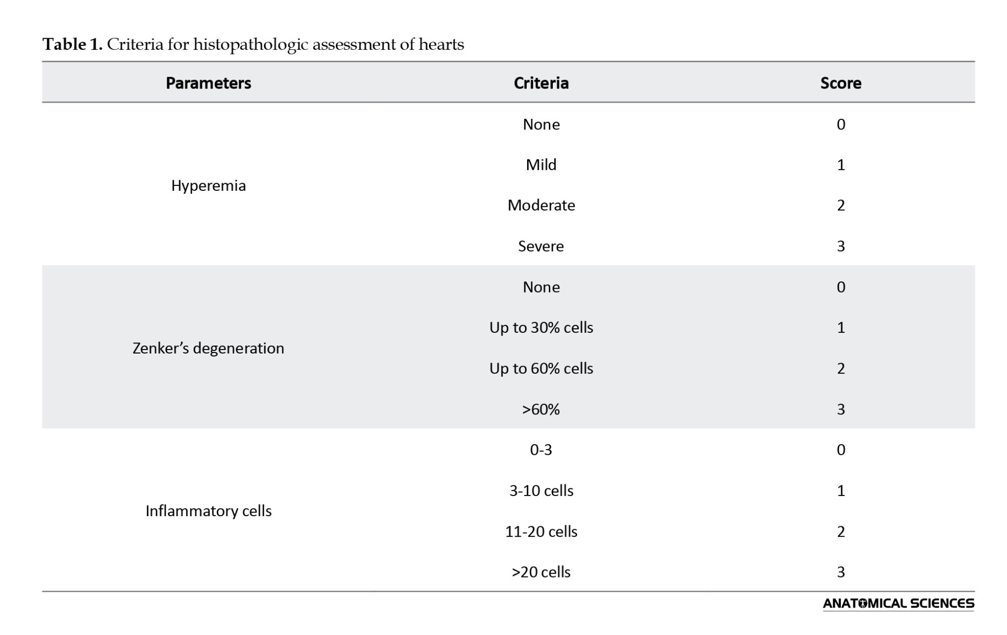
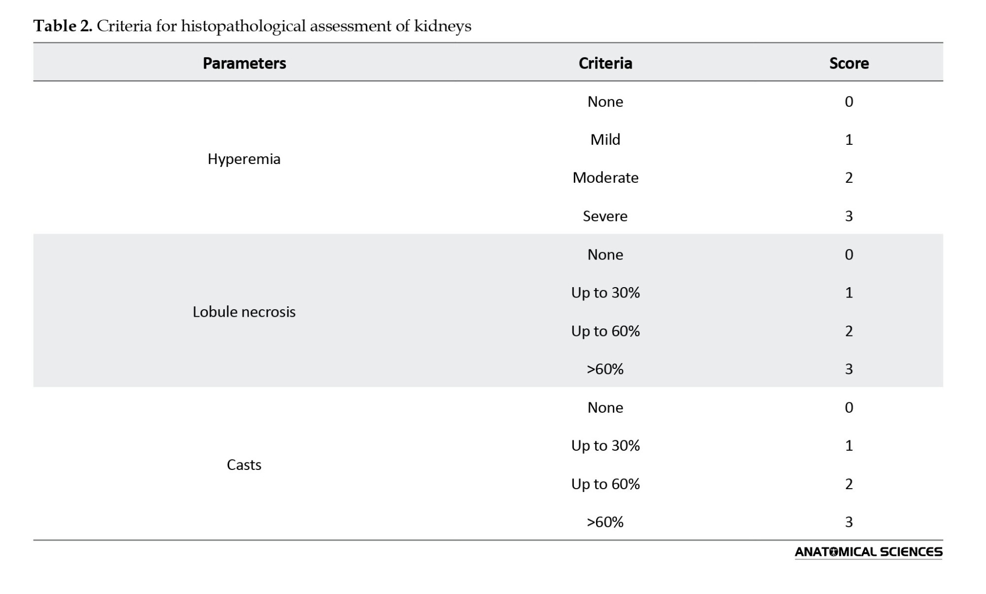
The tissue samples were cut at 3-5 μm, mounted on slides, and stained with hematoxylin and eosin (H&E). Each slide was then observed under a light microscope with 200x magnification across an average of 5 fields. They were then scored according to the criteria mentioned in Table 1 for hearts and Table 2 for kidneys [11]. For each slide, the minimum score was zero, and the maximum was 9. Higher scores would indicate higher degrees of tissue damage.
Statistical analysis
Categorical variables were expressed as frequency (percentage) and continuous data were expressed as median (interquartile range). All statistical analyses were performed in SPSS software, version 24. Data were analyzed using one-way analysis of variance (ANOVA) and Tukey’s test for blood parameters, as well as Kruskal-Wallis and Mann-Whitney U tests for the histopathological study. P<0.05 was considered as statistically significant.
Results
All mice survived during the 10-day study period and successfully reached the necropsy stage.
Total antioxidant capacity
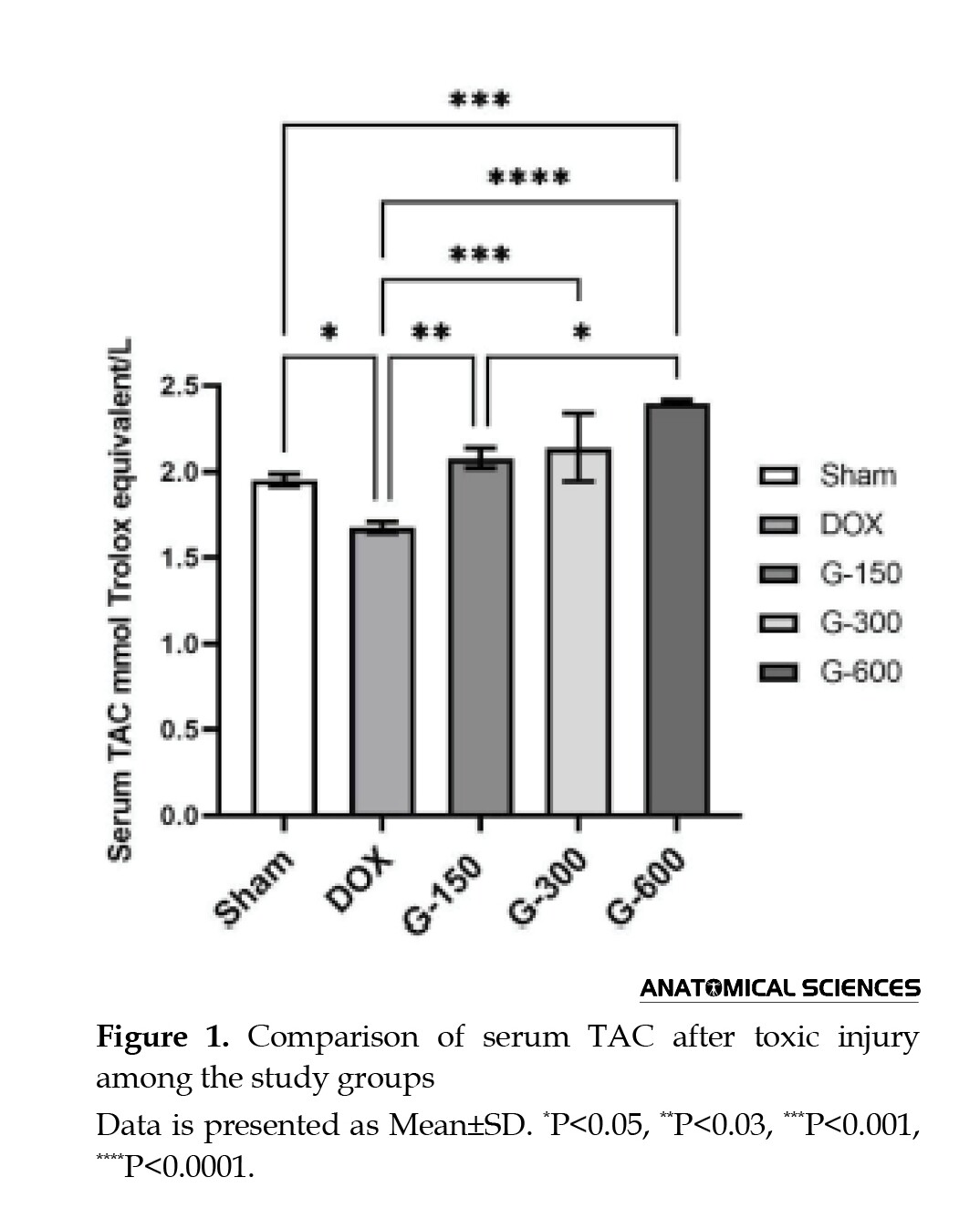
In Figure 1, the comparison of TAC between the study groups and the test results are illustrated. Group 5, treated with 600 mg/kg AGE, showed the highest TAC, followed by groups 4 and 3. The TAC in the control group was significantly higher than in the DOX group (P<0.05). There was a statistically significant difference in the TAC of groups 4 and 5 compared to the DOX group (P<0.0001). These results indicate the dose-dependent antioxidant effect of AGE in the body.
TOS
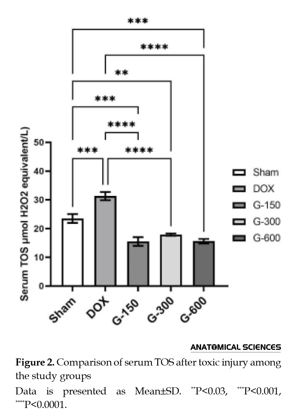
In Figure 2, the comparison of TOS between the study groups and the test results are illustrated. As can be seen, the difference among the groups was statistically significant.
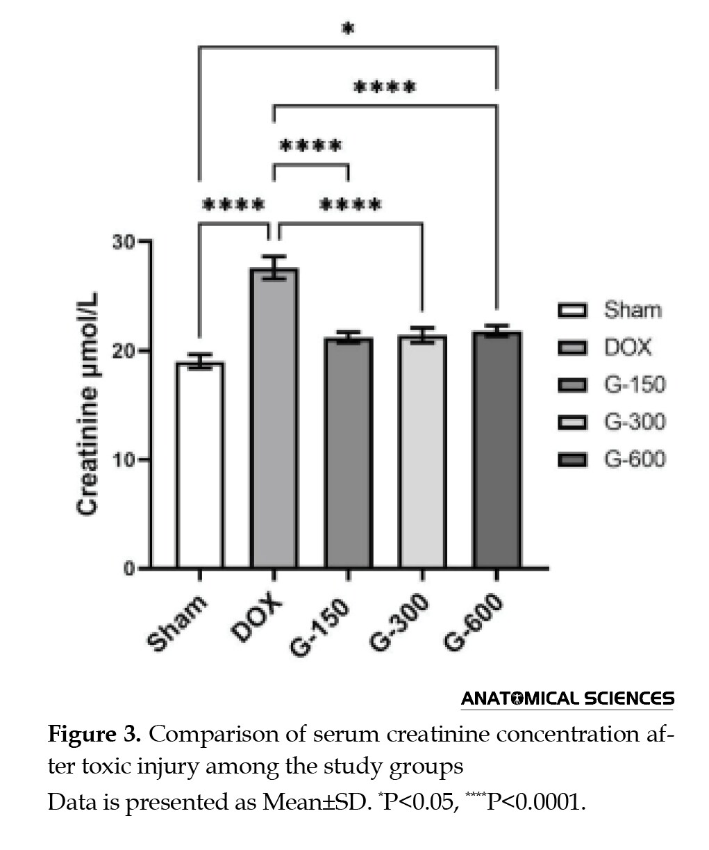
Serum creatinine concentration
As can be seen in Figure 3, there was a significantly higher serum creatinine concentration in the DOX group compared to the control group (P<0.0001). Moreover, all garlic extract groups showed a significantly lower serum creatinine concentration compared the DOX group (P<0.0001). Group 5 (received the highest dose of AGE) had a significantly different serum creatinine concentration compared to both the DOX group and the control group. The 600 mg/kg dose of AGE had a weaker protective effect on the kidneys.
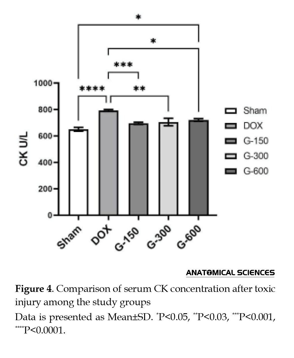
Serum creatine kinase concentration
According to Figure 4, the serum CK concentration was significantly different in the control group and in all garlic extract groups compared to the DOX group. This indicates the destructive effect of the drug on heart tissue and sheds insight into the protective effect of different doses of garlic extract. On the other hand, the 300 mg/kg and 600 mg/kg doses of AGE had a less significant difference than the 150 mg/kg dose compared to the DOX group, indicating that the 150 mg/kg dose is considerably better than the other two doses.
Histopathological changes in the heart
In Figure 5, the mean scores of histopathological changes in the heart of study groups and the results of Kruskal-Wallis and Mann-Whitney tests for their comparison are presented.
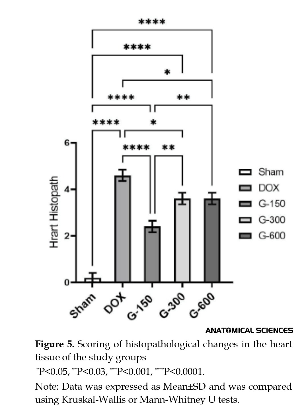
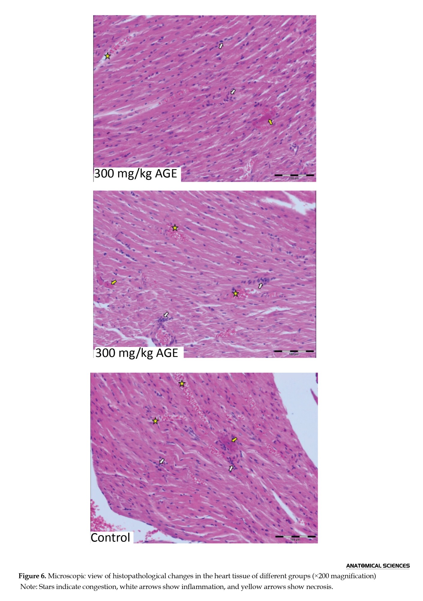
Figure 6 provides microscopic images of these changes. As can be seen, the control group was completely healthy and no inflammation was observed, while there were significant differences between the DOX and the AGE groups (P<0.0001). DOX caused changes in the heart tissue, including necrosis, inflammation, and congestion.
The DOX and the AGE groups had significant differences in terms of histopathological changes in the heart compared to the control group. There was no significant difference between groups 4 and 5 (P>0.05). Among the AGE groups, group 3 had the least tissue changes recorded, with a significant difference compared to groups 4 and 5 (P<0.01). The findings suggest that AGE has a protective effect on the cardiac tissue of mice with DOX-induced toxicity, and that the 150 mg/kg dose of AGE has a better protective effect against cardiac lesions.
Histopathological changes in the kidneys
Figure 7 presents the mean scores of histopathological changes in the kidneys of the study groups and the results of Kruskal-Wallis and Mann-Whitney tests for their comparison.
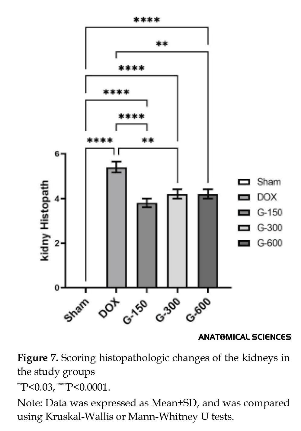
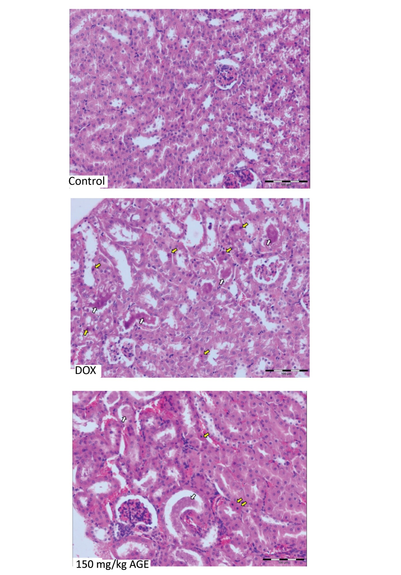
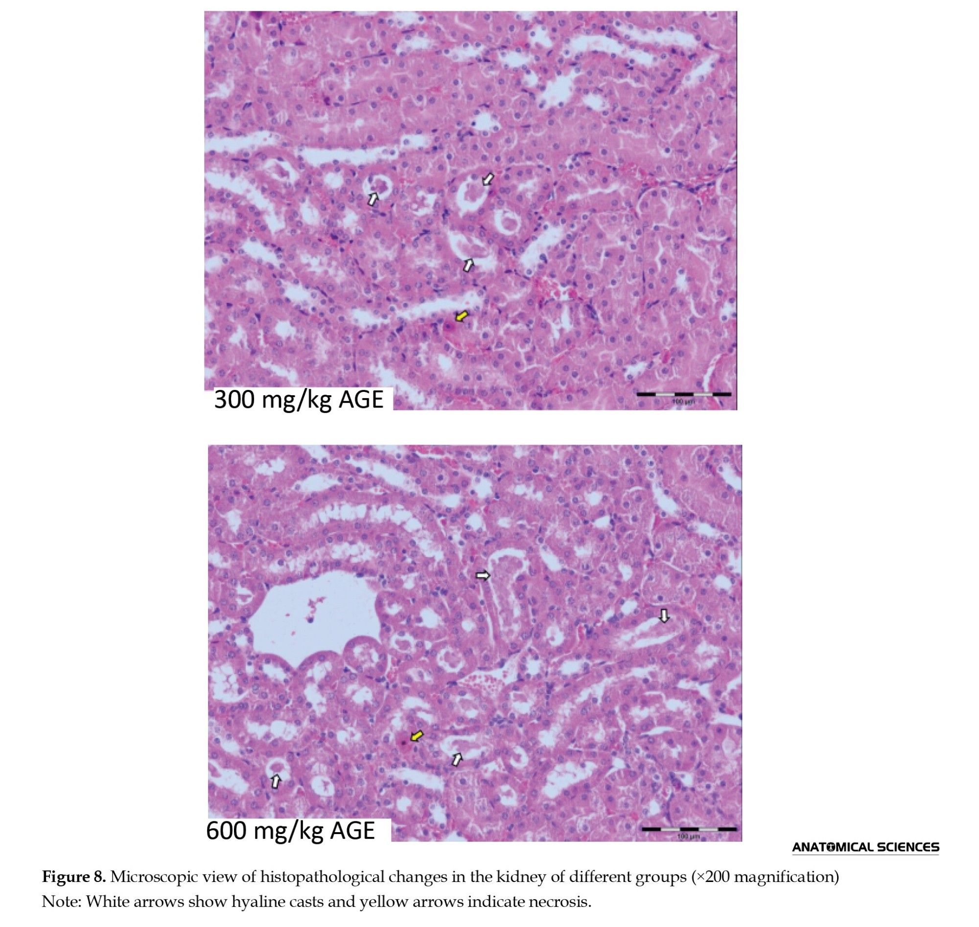
Figure 8 provides microscopic images of these changes. As can be seen, the control group exhibited normal structures and healthy tubules. The DOX group had the highest levels of necrosis and casts and a significant amount of edema compared to other groups (P<0.0001). All AGE groups had significant differences compared to the DOX group (P<0.0001). Group 3 (received 150 mg/kg AGE) did not show a significant difference compared to groups 4 and 5 (received 300 or 600 mg/kg AGE).
Discussion
In this study, the injection of a single dose of DOX (15 mg/kg) to female mice caused significant decline in all study outcomes. However, treatment with different doses of AGE showed significant improvement, where, the 150 mg/kg dose had the highest significant effect, except on TAC (where it was determined that there was a dose-dependent relationship between TAC and AGE dose) and histopathological changes in the kidney (where all treatment groups had similar scores).
Based on the histopathological examinations of the heart tissue, the non-treated group (DOX group) showed significant inflammation, necrosis, or casts/edema while the treatment groups showed much better results, where the group treated with 150 mg/kg AGE had the best results, indicating that this dose had the best protective effect compared to 300 mg/kg or 600 mg/kg doses. These results are consistent with existing literature that DOX causes both cardiac and generalized damage. The mechanism of the mentioned damages has been investigated in many studies [1, 12, 13].
The protective effect of AGE on DOX-induced toxicity has also been investigated in other studies. A 2010 study by Alkreathy et al. also showed that AGE is potentially protective against DOX-induced cardiotoxicity. Their study had longer duration, used different dose of AGE (250 mg/kg) and DOX (25 mg/kg) doses, and had different study outcomes [13]. In a 2009 study with similar parameters over 40 days, only 3 out of 10 mice from the DOX group survived. The electron micrograph assessment of the heart with DOX-induced toxicity showed vacuolization of muscle cells, disrupted myofibrils, and swollen mitochondria. On the other hand, the mice treated with AGE showed no serious lesions in the heart muscle cells [14].
The proposed mechanisms for such effects of GAE can be related to different antioxidant compounds of the garlic extract including SAC, which has been reported to have antioxidant, anti-cancer, and hepatoprotective activities [15, 16], and allicin, which has the power to prevent apoptosis by reducing levels of Bax gene expression and cytochrome C released from mitochondria [17]. It has been shown that after induction of toxicity by DOX, the expression of the Bax gene increases, the expression of the Bcl-2 gene decreases [18], mitochondrial membrane stability decreases, and permeability to cytochrome C is enhanced. Cytochrome C plays a direct role in apoptosis [19]. Thus, it can be said that the toxicity induced by DOX plays a role in mitochondrial membrane damage, but AGE disrupts this effect [20]. DOX-induced nephropathy is a common animal model for evaluating the therapeutic effects of compounds in human nephrotic syndrome. A study in 2020 showed that, after DOX administration, serum creatinine levels increased by up to 1.6 mg/dL in 26% of cats. The progressive kidney failure was reported as the cause of death in 7% of cats. A progressive increase in serum creatinine concentration was common from the beginning in cats receiving DOX [21]
In our study, the results of both histopathology examination and the serum creatinine concentration showed that all doses of AGE had similar protective effect on kidneys. Literature suggests that several phytochemicals demonstrate nuclear factor E2-related factor 2 (Nrf2)-activating properties and improve DOX-induced nephropathy. Tsai et al. in 2011 demonstrated that antroquinonol, an antioxidant derived from the mushroom Antrodia camphorate, has inhibitory effects on nitric oxide production and inflammatory reactions, and successfully protects mice kidneys from DOX-induced nephropathy. They suggested that the increase in renal Nrf2 and GSH peroxidase activity, along with inhibition of renal nuclear factor-κB (NF-κB) and lower levels of transforming growth factor (TGF)-β1, played a role in this protective effect [22]. This is line with our study because AGE and Antroq may exert their protective effects through similar pathways.
Overall, with the observed increase in TAC, and decrease in TOC and cardiac and kidney damage in groups treated with AGE, we can say that AGE has an attenuating effect on DOX-induced oxidative stress. However, a question may be raised: Does AGE simultaneously reduce the anti-tumoral and cytotoxic activity caused by DOX? In a 2011 study on tumor-bearing mice, it was found that AGE not only did not interrupt DOX-induced cytotoxic activity, but also enhanced it. The mean survival of mice with DOX use increased from 50 to 88 days after administration of AGE [23]. This effect may be due to the suppression of p-glycoprotein-associated energy-dependent efflux of the DOX pump, leading to a higher intracellular drug concentration and increased cellular toxicity [24]. Moreover, AGE is one of many herbal antioxidants that can potentially benefit patients undergoing chemotherapy. Hence, there is a need for more comprehensive research in this area, comparing the diverse effects of each herbal medicine. One study in 2009 by Demirkaya et al. on the protective effects of different doses of proanthocyanidin, hazelnut, and garlic against DOX-induced injuries showed that only garlic caused a significant reduction in cardiac damage at high doses. Although the cardiac damage scores in the proanthocyanidin and high-dose hazelnut groups were lower than in the control group, the differences were not statistically significant [25].
One important limitation of this study was the fact that the mice were previously healthy, and our study period was only 10 days. Further studies with longer duration and with the inclusion of tumor-bearing mice or other chemotherapy agents, can provide much more concrete information on AGE’s protective effect against DOX-inducted toxicity.
Conclusion
It can be concluded that AGE can reduce the severity of toxic injuries in the heart and kidneys caused by DOX. Different doses of AGE have different effects. More studies are needed on the protective effects and potential side effects of each dose, and assessing whether or not these results are replicable in human models. Researchers should continue to identify the potential characteristics of toxic injuries caused by DOX and validate new herbal treatment methods for reducing these toxic injuries.
Ethical Considerations
Compliance with ethical guidelines
There were no ethical considerations to be considered in this research.
Funding
This research did not receive any grant from funding agencies in the public, commercial, or non-profit sectors.
Authors' contributions
All authors contributed equally to the conception and design of the study, data collection and analysis, interception of the results and drafting of the manuscript. Each author approved the final version of the manuscript for submission.
Conflict of interest
The authors declared no conflict of interest.
References
Doxorubicin, also known as Adriamycin, is a cytotoxic anthracycline drug that is most commonly used for treating human solid tumors and leukemias. However, its use is restricted due to its dose-dependent toxicity for multiple organs, especially the heart, lungs, and kidneys. Doxorubicin (DOX) induces cardiac injury through many mechanisms, including mitochondrial damage, buildup of reactive oxygen species (ROS), and damage to DNA, lipids, and proteins. Mitochondrial damage is known to occur a few hours after the DOX use [1]. The occurrence of this phenomenon is related to many factors, such as dose, age, gender, genetics, duration of treatment, pre-existing disorders, and the combination of cancer treatments [2]. In a retrospective analysis of over 4000 patients receiving DOX performed by Von Hoff et al. [3], 2.2% of the patients developed clinical signs and symptoms of congestive heart failure. Thus, there have been significant efforts to mitigate DOX-induced cardiotoxicity. Many strategies have been proposed, including either structural modification or intervention with chemical and natural products. Several products have been produced and tested in many experimental animal models to assess their effect on DOX-induced toxicity without reducing its therapeutic efficacy. Natural plant antioxidants are among such products.
Garlic is one of the most widely used medicinal plants worldwide. Its activity is mostly attributed to organosulfur compounds. When extracted and isolated, these compounds exhibit various useful properties, including but not limited to anti-oxidant, anti-inflammatory, and anti-apoptotic activity. Its key compounds include Allicin, diallyl trisulfide (DATS), L-cysteine, alliin, S-allylcysteine (SAC), and ajoene [4, 5]. Many garlic preparations exist, including liquid (aqueous, oil, or solvent extracts) and solid (dried powder and fresh garlic). Aged garlic extract (AGE) is among the most well-known garlic preparations [6, 7]. It is prepared by immersing raw garlic slices in a low ethanol concentration for about 12 months, kept at room temperature. This preparation results in an odorless extract that contains high concentrations of SAC, DATS, and alliin [7]. It has higher antioxidant potential than other garlic products, mainly due to its concentration of water-soluble organosulfur compounds. These compounds can neutralize ROS and reactive nitrogen species. Through this, AGE can protect lipids, proteins, and DNA from oxidation. Additionally, it increases glutathione (GSH) levels, protecting cells against oxidative stress [7, 8]. This study aims to investigate the potential protective effects of AGE against cardiac and renal toxicity caused by DOX in female mice.
Materials and Methods
Garlic extract preparation
Fresh garlic was purchased from a local market, cleaned, peeled, and then dried at room temperature (25 °C). Ten grams of dried garlic was added to 100 mL of a mix of 85% ethanol and distilled water. The mixture was gently stirred on an extractor (Soxhlet) for 72 hours to ensure proper extraction. The resulting solution was then filtered to obtain the primary extract. The primary extract was then placed in a vacuum distillation device, and at a temperature of 80 °C, the solvent was gradually evaporated over an hour, yielding the concentrated extract. This method effectively replicates the traditional creation of AGE, which takes over 12 months [6]. It was then stored in the freezer at -20 °C away from light for use in subsequent stages of the study.
Animals
Twenty-five healthy female BALB/c mice weighing about 20 grams were obtained from the Pasteur Institute of Iran. Their environments were identical and their handling complied with ethical standards. They were kept in standard cages with filtered water and adequate food at room temperature and a light/dark cycle of 12 hours. They were later randomly assigned to five groups of 5 as follows:
Group 1: As a control group, it received 2 mg/kg gavage of normal saline solution daily for 10 days;
Group 2: Named as DOX group, it received 2 mg/kg normal saline for 10 days plus a single intraperitoneal injection of 15 mg/kg DOX (Aldrich-Sigma Co.) on day 7.
Group 3: This group received DOX 15 mg/kg on day 7 plus the gavage of 150 mg/kg AGE for 10 days.
Group 4: This group received DOX 15 mg/kg on day 7 plus the gavage of 300 mg/kg AGE for 10 days.
Group 5: This group received DOX 15 mg/kg on day 7 plus a gavage of 600 mg/kg AGE for 10 days.
Study protocol and assess
The study took place over 10 consecutive days. At the end of 10 days, the mice were euthanized under deep anesthesia with ketamine, and blood samples were collected directly from their hearts and sent for biochemical analyses. Abdomens were cut open and hearts and kidneys were rapidly removed. Half of the recovered samples were then processed and fixed in a formalin solution for later inspection with a light microscope. The remaining half were stored in -20 °C for later analysis.
Biochemical study
Biochemical study included the assessment of serum creatinine concentration (To measure kidney function), serum creatine kinase (CK) concentration (as a biochemical proxy of cardiac muscle function), total antioxidant capacity (TAC), and total oxidant status (TOS).
The TAC was calculated using the ferric reducing/antioxidant power (FRAP) assay. This method is based on the serum’s ability to reduce ferric ions (+3) to ferrous ions (+2) and is conducted in the presence of a reagent called 2,4,6-Tri-(2-pyridyl)-s-triazin (TPTZ). Ferrous ions (Fe²⁺) react to TPTZ and form the blue Fe²⁺-TPTZ complex. This complex displays a maximum optical absorption at a wavelength of 593 nanometers, which was measured by a spectrophotometer (UNICO 2150-UV, China) [9].
To measure TOS, The Erel Technique was used. This method is based on the oxidation of ferrous (Fe²⁺) to ferric (Fe³⁺) ions and the formation of a complex in the presence of xylenol orange. The number of oxidizing molecules is directly related to the color intensity of the complex in the acidic solution. This intensity was measured by a spectrophotometer at a wavelength of 560 nanometers [10].
Histopathological study
The tissue samples were cut at 3-5 μm, mounted on slides, and stained with hematoxylin and eosin (H&E). Each slide was then observed under a light microscope with 200x magnification across an average of 5 fields. They were then scored according to the criteria mentioned in Table 1 for hearts and Table 2 for kidneys [11]. For each slide, the minimum score was zero, and the maximum was 9. Higher scores would indicate higher degrees of tissue damage.
Statistical analysis
Categorical variables were expressed as frequency (percentage) and continuous data were expressed as median (interquartile range). All statistical analyses were performed in SPSS software, version 24. Data were analyzed using one-way analysis of variance (ANOVA) and Tukey’s test for blood parameters, as well as Kruskal-Wallis and Mann-Whitney U tests for the histopathological study. P<0.05 was considered as statistically significant.
Results
All mice survived during the 10-day study period and successfully reached the necropsy stage.
Total antioxidant capacity
In Figure 1, the comparison of TAC between the study groups and the test results are illustrated. Group 5, treated with 600 mg/kg AGE, showed the highest TAC, followed by groups 4 and 3. The TAC in the control group was significantly higher than in the DOX group (P<0.05). There was a statistically significant difference in the TAC of groups 4 and 5 compared to the DOX group (P<0.0001). These results indicate the dose-dependent antioxidant effect of AGE in the body.
TOS
In Figure 2, the comparison of TOS between the study groups and the test results are illustrated. As can be seen, the difference among the groups was statistically significant.
Serum creatinine concentration
As can be seen in Figure 3, there was a significantly higher serum creatinine concentration in the DOX group compared to the control group (P<0.0001). Moreover, all garlic extract groups showed a significantly lower serum creatinine concentration compared the DOX group (P<0.0001). Group 5 (received the highest dose of AGE) had a significantly different serum creatinine concentration compared to both the DOX group and the control group. The 600 mg/kg dose of AGE had a weaker protective effect on the kidneys.
Serum creatine kinase concentration
According to Figure 4, the serum CK concentration was significantly different in the control group and in all garlic extract groups compared to the DOX group. This indicates the destructive effect of the drug on heart tissue and sheds insight into the protective effect of different doses of garlic extract. On the other hand, the 300 mg/kg and 600 mg/kg doses of AGE had a less significant difference than the 150 mg/kg dose compared to the DOX group, indicating that the 150 mg/kg dose is considerably better than the other two doses.
Histopathological changes in the heart
In Figure 5, the mean scores of histopathological changes in the heart of study groups and the results of Kruskal-Wallis and Mann-Whitney tests for their comparison are presented.
Figure 6 provides microscopic images of these changes. As can be seen, the control group was completely healthy and no inflammation was observed, while there were significant differences between the DOX and the AGE groups (P<0.0001). DOX caused changes in the heart tissue, including necrosis, inflammation, and congestion.
The DOX and the AGE groups had significant differences in terms of histopathological changes in the heart compared to the control group. There was no significant difference between groups 4 and 5 (P>0.05). Among the AGE groups, group 3 had the least tissue changes recorded, with a significant difference compared to groups 4 and 5 (P<0.01). The findings suggest that AGE has a protective effect on the cardiac tissue of mice with DOX-induced toxicity, and that the 150 mg/kg dose of AGE has a better protective effect against cardiac lesions.
Histopathological changes in the kidneys
Figure 7 presents the mean scores of histopathological changes in the kidneys of the study groups and the results of Kruskal-Wallis and Mann-Whitney tests for their comparison.
Figure 8 provides microscopic images of these changes. As can be seen, the control group exhibited normal structures and healthy tubules. The DOX group had the highest levels of necrosis and casts and a significant amount of edema compared to other groups (P<0.0001). All AGE groups had significant differences compared to the DOX group (P<0.0001). Group 3 (received 150 mg/kg AGE) did not show a significant difference compared to groups 4 and 5 (received 300 or 600 mg/kg AGE).
Discussion
In this study, the injection of a single dose of DOX (15 mg/kg) to female mice caused significant decline in all study outcomes. However, treatment with different doses of AGE showed significant improvement, where, the 150 mg/kg dose had the highest significant effect, except on TAC (where it was determined that there was a dose-dependent relationship between TAC and AGE dose) and histopathological changes in the kidney (where all treatment groups had similar scores).
Based on the histopathological examinations of the heart tissue, the non-treated group (DOX group) showed significant inflammation, necrosis, or casts/edema while the treatment groups showed much better results, where the group treated with 150 mg/kg AGE had the best results, indicating that this dose had the best protective effect compared to 300 mg/kg or 600 mg/kg doses. These results are consistent with existing literature that DOX causes both cardiac and generalized damage. The mechanism of the mentioned damages has been investigated in many studies [1, 12, 13].
The protective effect of AGE on DOX-induced toxicity has also been investigated in other studies. A 2010 study by Alkreathy et al. also showed that AGE is potentially protective against DOX-induced cardiotoxicity. Their study had longer duration, used different dose of AGE (250 mg/kg) and DOX (25 mg/kg) doses, and had different study outcomes [13]. In a 2009 study with similar parameters over 40 days, only 3 out of 10 mice from the DOX group survived. The electron micrograph assessment of the heart with DOX-induced toxicity showed vacuolization of muscle cells, disrupted myofibrils, and swollen mitochondria. On the other hand, the mice treated with AGE showed no serious lesions in the heart muscle cells [14].
The proposed mechanisms for such effects of GAE can be related to different antioxidant compounds of the garlic extract including SAC, which has been reported to have antioxidant, anti-cancer, and hepatoprotective activities [15, 16], and allicin, which has the power to prevent apoptosis by reducing levels of Bax gene expression and cytochrome C released from mitochondria [17]. It has been shown that after induction of toxicity by DOX, the expression of the Bax gene increases, the expression of the Bcl-2 gene decreases [18], mitochondrial membrane stability decreases, and permeability to cytochrome C is enhanced. Cytochrome C plays a direct role in apoptosis [19]. Thus, it can be said that the toxicity induced by DOX plays a role in mitochondrial membrane damage, but AGE disrupts this effect [20]. DOX-induced nephropathy is a common animal model for evaluating the therapeutic effects of compounds in human nephrotic syndrome. A study in 2020 showed that, after DOX administration, serum creatinine levels increased by up to 1.6 mg/dL in 26% of cats. The progressive kidney failure was reported as the cause of death in 7% of cats. A progressive increase in serum creatinine concentration was common from the beginning in cats receiving DOX [21]
In our study, the results of both histopathology examination and the serum creatinine concentration showed that all doses of AGE had similar protective effect on kidneys. Literature suggests that several phytochemicals demonstrate nuclear factor E2-related factor 2 (Nrf2)-activating properties and improve DOX-induced nephropathy. Tsai et al. in 2011 demonstrated that antroquinonol, an antioxidant derived from the mushroom Antrodia camphorate, has inhibitory effects on nitric oxide production and inflammatory reactions, and successfully protects mice kidneys from DOX-induced nephropathy. They suggested that the increase in renal Nrf2 and GSH peroxidase activity, along with inhibition of renal nuclear factor-κB (NF-κB) and lower levels of transforming growth factor (TGF)-β1, played a role in this protective effect [22]. This is line with our study because AGE and Antroq may exert their protective effects through similar pathways.
Overall, with the observed increase in TAC, and decrease in TOC and cardiac and kidney damage in groups treated with AGE, we can say that AGE has an attenuating effect on DOX-induced oxidative stress. However, a question may be raised: Does AGE simultaneously reduce the anti-tumoral and cytotoxic activity caused by DOX? In a 2011 study on tumor-bearing mice, it was found that AGE not only did not interrupt DOX-induced cytotoxic activity, but also enhanced it. The mean survival of mice with DOX use increased from 50 to 88 days after administration of AGE [23]. This effect may be due to the suppression of p-glycoprotein-associated energy-dependent efflux of the DOX pump, leading to a higher intracellular drug concentration and increased cellular toxicity [24]. Moreover, AGE is one of many herbal antioxidants that can potentially benefit patients undergoing chemotherapy. Hence, there is a need for more comprehensive research in this area, comparing the diverse effects of each herbal medicine. One study in 2009 by Demirkaya et al. on the protective effects of different doses of proanthocyanidin, hazelnut, and garlic against DOX-induced injuries showed that only garlic caused a significant reduction in cardiac damage at high doses. Although the cardiac damage scores in the proanthocyanidin and high-dose hazelnut groups were lower than in the control group, the differences were not statistically significant [25].
One important limitation of this study was the fact that the mice were previously healthy, and our study period was only 10 days. Further studies with longer duration and with the inclusion of tumor-bearing mice or other chemotherapy agents, can provide much more concrete information on AGE’s protective effect against DOX-inducted toxicity.
Conclusion
It can be concluded that AGE can reduce the severity of toxic injuries in the heart and kidneys caused by DOX. Different doses of AGE have different effects. More studies are needed on the protective effects and potential side effects of each dose, and assessing whether or not these results are replicable in human models. Researchers should continue to identify the potential characteristics of toxic injuries caused by DOX and validate new herbal treatment methods for reducing these toxic injuries.
Ethical Considerations
Compliance with ethical guidelines
There were no ethical considerations to be considered in this research.
Funding
This research did not receive any grant from funding agencies in the public, commercial, or non-profit sectors.
Authors' contributions
All authors contributed equally to the conception and design of the study, data collection and analysis, interception of the results and drafting of the manuscript. Each author approved the final version of the manuscript for submission.
Conflict of interest
The authors declared no conflict of interest.
References
- Song L, Qiu Q, Ju F, Zheng C. Mechanisms of doxorubicin-induced cardiac inflammation and fibrosis; therapeutic targets and approaches. Archives of Biochemistry and Biophysics. 2024; 761:110140. [DOI:10.1016/j.abb.2024.110140] [PMID]
- Belger C, Abrahams CB, Imamdin A, Lecour S. Doxorubicin-induced cardiotoxicity and risk factors. J Cardiol Heart Vasc. 2023; 50:101332. [DOI:10.1016/j.ijcha.2023.101332] [PMID]
- Von Hoff DD, Layard MW, Basa P, Davis HL Jr, Von Hoff AL, Rozencweig M, et al. Risk factors for doxorubicin-induced congestive heart failure. Annals of Internal Medicine. 1979; 91(5):710-7. [DOI:10.7326/0003-4819-91-5-710] [PMID]
- Abdel-Daim MM, Kilany OE, Khalifa HA, Ahmed AAM. Allicin ameliorates doxorubicin-induced cardiotoxicity in rats via suppression of oxidative stress, inflammation and apoptosis. Cancer Chemotherapy and Pharmacology. 2017; 80(4):745-53. [DOI:10.1007/s00280-017-3413-7] [PMID]
- Novakovic J, Muric M, Bradic J, Ramenskaya G, Jakovljevic V, Jeremic N. Diallyl trisulfide and cardiovascular health: evidence and potential molecular mechanisms. International Journal of Molecular Sciences. 2024; 25(18):9831. [DOI:10.3390/ijms25189831] [PMID]
- Arreola R, Quintero-Fabián S, López-Roa RI, Flores-Gutiérrez EO, Reyes-Grajeda JP, Carrera-Quintanar L, et al. Immunomodulation and anti-inflammatory effects of garlic compounds. Journal of Immunology Research. 2015; 2015:401630. [DOI:10.1155/2015/401630] [PMID]
- Borek C. Antioxidant health effects of aged garlic extract. The Journal of Nutrition. 2001; 131(3s):1010S-5S. [DOI:10.1093/jn/131.3.1010S] [PMID]
- Jeong YY, Ryu JH, Shin JH, Kang MJ, Kang JR, Han J, et al. Comparison of anti-oxidant and anti-inflammatory effects between fresh and aged black garlic extracts. Molecules. 2016; 21(4):430. [DOI:10.3390/molecules21040430] [PMID]
- Benzie IF, Strain JJ. The ferric reducing ability of plasma (FRAP) as a measure of “antioxidant power”: The FRAP assay. Analytical Biochemistry. 1996; 239(1):70-6. [DOI:10.1006/abio.1996.0292] [PMID]
- Erel O. A new automated colorimetric method for measuring total oxidant status. Clinical Biochemistry. 2005; 38(12):1103-11. [DOI:10.1016/j.clinbiochem.2005.08.008] [PMID]
- Demir F, Güzel A, Kat C, Karadeniz C, Akdemir U, Okuyucu A, et al. A combination of methylprednisolone and quercetin is effective for the treatment of cardiac contusion following blunt chest trauma in rats. Brazilian Journal of Medical and Biological Research. 2014; 47(9):766-72. [DOI:10.1590/1414-431X20144021] [PMID]
- Van Vleet JF, Ferrans VJ, Weirich WE. Cardiac disease induced by chronic adriamycin administration in dogs and an evaluation of vitamin E and selenium as cardioprotectants. American Journal of Pathology. 1980; 99(1):13-42. [PMID]
- Alkreathy H, Damanhouri ZA, Ahmed N, Slevin M, Osman AM. Aged garlic extract protects against doxorubicin-induced cardiotoxicity in rats. Food and Chemical Toxicology. 2010; 48(3):951-956. [DOI:10.1016/j.fct.2010.01.005] [PMID]
- Libero ML, Montero-Hidalgo AJ, Recinella L, Luque RM, Generali D, Acquaviva A, et al. The protective effects of an aged black garlic water extract on the prostate. Nutrients. 2024; 16(17):3025. [DOI:10.3390/nu16173025] [PMID]
- Alkreathy HM, Damanhouri ZA, Ahmed N, Slevin M, Osman AM. Mechanisms of cardioprotective effect of aged garlic extract against doxorubicin-induced cardiotoxicity. Integrative Cancer Therapies. 2012; 11(4):364-70. [DOI:10.1177/1534735411426726] [PMID]
- Al-Malky HS, Osman AM, Damanhouri ZA, Alkreathy HM, Al Aama JY, Ramadan WS, et al. Modulation of doxorubicin-induced expression of the multidrug resistance gene in breast cancer cells by diltiazem and protection against cardiotoxicity in experimental animals. Cancer Cell International. 2019; 19:191. [DOI:10.1186/s12935-019-0912-0] [PMID]
- Savairam VD, Patil NA, Borate SR, Ghaisas MM, Shete RV. Allicin: A review of its important pharmacological activities. Pharmacological Research - Modern Chinese Medicine. 2023; 100283. [DOI:10.1016/j.prmcm.2023.100283]
- Gorji SM, Malekshah AK. [Effect of doxorubicin on Bcl2 and Bax expression in rat heart (Persian)]. Journal of Gorgan University of Medical Sciences. 2013; 15:19-24. [Link]
- Skulachev VP. Cytochrome c in the apoptotic and antioxidant cascades. FEBS Letters. 1998; 423(3):275-80. [DOI:10.1016/S0014-5793(98)00061-1] [PMID]
- Chen RC, Xu XD, Liu XZ, Sun GB, Zhu YD, Dong X, et al. Total flavonoids from Clinopodium chinense (Benth.) O. Ktze protect against doxorubicin-induced cardiotoxicity in vitro and in vivo. Evid Based Complement Alternat Med. 2015; 2015:472565. [DOI:10.1155/2015/472565] [PMID]
- Kopecny L, Palm CA, Skorupski KA, Delgado M, Rebhun RB. Risk factors associated with progressive increases in serum creatinine concentrations in cats with cancer receiving doxorubicin. Journal of Veterinary Internal Medicine. 2020; 34(5):2048-55. [DOI:10.1111/jvim.15867] [PMID]
- Tsai PY, Ka SM, Chao TK, Chang JM, Lin SH, Li CY, et al. Antroquinonol reduces oxidative stress by enhancing the Nrf2 signaling pathway and inhibits inflammation and sclerosis in focal segmental glomerulosclerosis mice. Free Radical Biology and Medicine. 2011; 50(11):1503-16. [DOI:10.1016/j.freeradbiomed.2011.02.029] [PMID]
- Colín-González AL, Santana RA, Silva-Islas CA, Chánez-Cárdenas ME, Santamaría A, Maldonado PD. The antioxidant mechanisms underlying the aged garlic extract- and S-allylcysteine-induced protection. Oxid Med Cell Longev. 2012; 2012:907162. [DOI:10.1155/2012/907162] [PMID]
- Rais N, Akash V, Rizwan A, Kumar M, Radha D, Chandran D, et al. S-allyl-l-cysteine - a garlic bioactive: physicochemical nature, mechanism, pharmacokinetics, and health-promoting activities. Journal of Functional Foods. 2023; 105657. [DOI:10.1016/j.jff.2023.105657]
- Demirkaya E, Avci A, Kesik V, Karslioglu Y, Oztas E, Kismet E, et al. Cardioprotective roles of aged garlic extract, grape seed proanthocyanidin, and hazelnut on doxorubicin-induced cardiotoxicity. Canadian Journal of Physiology and Pharmacology. 2009; 87(8):633-40. [Link]
Type of Study: Original |
Subject:
Histology
Received: 2025/01/27 | Accepted: 2025/02/16 | Published: 2023/08/30
Received: 2025/01/27 | Accepted: 2025/02/16 | Published: 2023/08/30
Send email to the article author
| Rights and permissions | |
 |
This work is licensed under a Creative Commons Attribution-NonCommercial 4.0 International License. |

Copyright © The Author(s);
This is an open access article distributed under the terms of the Creative Commons Attribution License (CC-By-NC), which permits use, distribution, and reproduction in any medium, provided the original work is properly cited and is not used for commercial purposes.
Contact Information
