Sat, Jul 25, 2026
Volume 14, Issue 2 (Summer & Autumn 2017)
ASJ 2017, 14(2): 97-100 |
Back to browse issues page

Download citation:
BibTeX | RIS | EndNote | Medlars | ProCite | Reference Manager | RefWorks
Send citation to:


BibTeX | RIS | EndNote | Medlars | ProCite | Reference Manager | RefWorks
Send citation to:
Eftekhar Vaghefi S H, Dehghani Soltani S, Babaee A. An Uncommon Anatomical Variation of the Sciatic Nerve. ASJ 2017; 14 (2) :97-100
URL: http://anatomyjournal.ir/article-1-161-en.html
URL: http://anatomyjournal.ir/article-1-161-en.html


1- Department of Anatomical Sciences, School of Medicine, Kerman University of Medical Sciences, Kerman, Iran.
Full-Text [PDF 450 kb]
(2080 Downloads)
| Abstract (HTML) (6663 Views)
Full-Text: (3402 Views)
1. Introduction
The sciatic nerve, also known as ischiatic nerve, is the largest nerve of the lumbosacral nervous plexus (L4-S3). This nerve enters the greater sciatic foramen and in order to arrive the gluteal area, passes through the inferior border of piriformis muscle [1]. Inferiorly, in a region between the greater trochanter and ischial tuberosity, the sciatic nerve is located deep to the gluteus maximus [2]. At the inferior margin of the quadratus femoris, sciatic nerve enters the back of the thigh. In this compartment, it is situated posteriorly to the adductor magnus overlapping by means of biceps femoris [2, 3].
Usually, in the lower third of thigh and sometimes within the gluteal region, this nerve is divided into 2 branches: the tibial and the common peroneal nerves, and innervates all muscles of the leg and foot via these branches [4, 5]. Therefore, the sciatic nerve palsy leads to severe difficulty in walking [6]. Anatomical variations of ischiatic nerve are very important to many surgical procedures [7]. Identification of such variations is essential to inhibit nerve injury and postoperative complications [8, 9]. Thus, the current study aimed at describing a thick septum situated posterior to the sciatic nerve.
2. Case Report
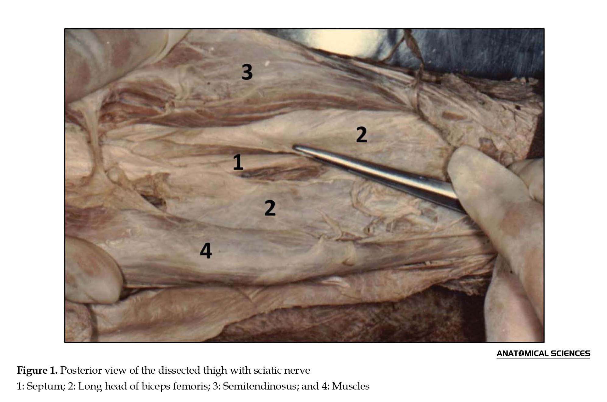
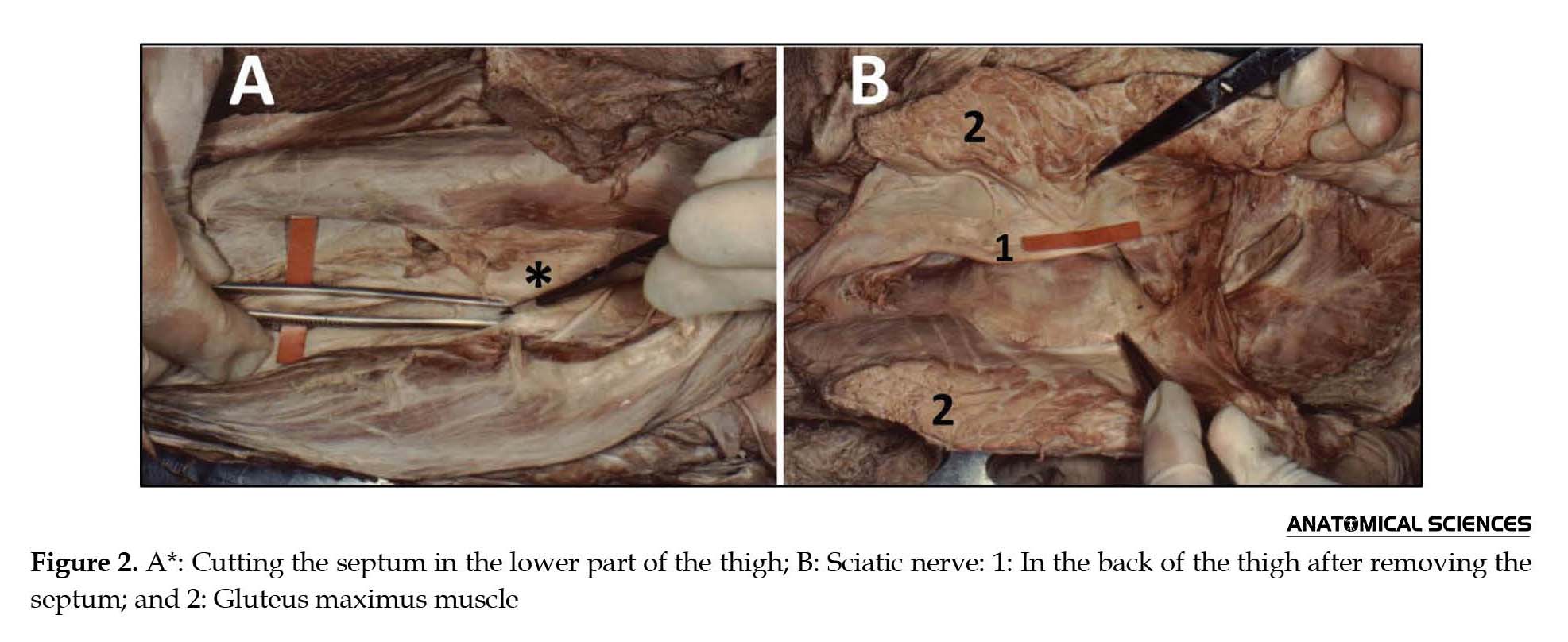
The current study was conducted in Kerman University of Medical Sciences, Kerman, Iran. During a cadaver (male, 35 to 40 year of age) dissection in anatomy learning, a rare variation was detected in the posterior region of the thigh. In this case, a thick white septum situated posterior to the sciatic nerve widespread throughout the back of thigh. Also, this septum (Figure 1) lay just deep to muscles such as semitendinosus, semimembranosus, and biceps femoris. This septum was detected after cleaning the connective tissue of this region; sciatic nerve and its 2 major divisions were found as soon as cutting the septum (Figure 2).
The sciatic nerve, also known as ischiatic nerve, is the largest nerve of the lumbosacral nervous plexus (L4-S3). This nerve enters the greater sciatic foramen and in order to arrive the gluteal area, passes through the inferior border of piriformis muscle [1]. Inferiorly, in a region between the greater trochanter and ischial tuberosity, the sciatic nerve is located deep to the gluteus maximus [2]. At the inferior margin of the quadratus femoris, sciatic nerve enters the back of the thigh. In this compartment, it is situated posteriorly to the adductor magnus overlapping by means of biceps femoris [2, 3].
Usually, in the lower third of thigh and sometimes within the gluteal region, this nerve is divided into 2 branches: the tibial and the common peroneal nerves, and innervates all muscles of the leg and foot via these branches [4, 5]. Therefore, the sciatic nerve palsy leads to severe difficulty in walking [6]. Anatomical variations of ischiatic nerve are very important to many surgical procedures [7]. Identification of such variations is essential to inhibit nerve injury and postoperative complications [8, 9]. Thus, the current study aimed at describing a thick septum situated posterior to the sciatic nerve.
2. Case Report
The current study was conducted in Kerman University of Medical Sciences, Kerman, Iran. During a cadaver (male, 35 to 40 year of age) dissection in anatomy learning, a rare variation was detected in the posterior region of the thigh. In this case, a thick white septum situated posterior to the sciatic nerve widespread throughout the back of thigh. Also, this septum (Figure 1) lay just deep to muscles such as semitendinosus, semimembranosus, and biceps femoris. This septum was detected after cleaning the connective tissue of this region; sciatic nerve and its 2 major divisions were found as soon as cutting the septum (Figure 2).
3. Discussion
The identification of anatomical variations in muscles, vessels, and nerves are very important to clinical practices [10-12]. During the development of human embryo, the nerves enter to the lower limb to forms the 2 main nervous plexuses of these region including lumbar and sacral plexuses. The ischiatic nerve as soon as differentiation from plexuses enters the gluteal area through the greater sciatic foramen [13, 14].
The first part of sciatic is about 2 centimeters wide. It is also the thickest as well as the greatest nerve in the human body [15]. Many variations in this nerve were reported so far [16, 17]. Nayak et al. described the trifurcation of this nerve including the common peroneal, lateral cutaneous, and tibial nerves [18]. Similar studies demonstrated that sciatic nerve at a varying level of the back of the thigh is divided into 2 branches. Usually, the site of the sciatic bifurcation is between the middle third and lower third of the femoral height, close the entrance of the popliteal fossa [1, 5, 19]. The sciatic nerve may pass through the piriformis muscle and lead to the sciatica [20, 21].
The common fibular nerve passing superficial to the superior gemellus and tibial nerve passing deep to this muscle were reported in the literature [22]. Ultrasound guidance is an advanced technique specified to nerve localization [23]. In the current study, a new anatomical variation of sciatic nerve was described for the first time. This variation is very important because it may produce interference at time of ultrasound guidance procedure for sciatic nerve. Thorough knowledge of this septum is essential to establish a diagnosis and perform surgery in the back region of the thigh. Anatomical variations in the origination, termination, and construction of the ischiatic nerve as well as its relationship with the muscles or septum are very important [24, 25]. The identification of such variations is necessary and vital for surgeons and clinicians while scheduling clinical interventions.
Acknowledgments
This article was financially supported by Kerman University of Medical Sciences.
Conflict of Interest
The authors declared no conflicts of interest.
References
- Kotian SR, Sinha A, Souza AS, Sumalatha S. Variations of the sciatic nerve and its relation with the piriformis muscle in South Indian population. Journal of Experimental and Integrative Medicine. 2015; 5(3):144-8. doi: 10.5455/jeim.200515.or.132
- Moore KL, Dalley AF, Agur AM. Clinically oriented anatomy. Philadelphia: Lippincott Williams & Wilkins; 2013.
- Güvençer M, Akyer P, Iyem C, Tetik S, Naderi S. Anatomic considerations and the relationship between the piriformis muscle and the sciatic nerve. Surgical and Radiologic Anatomy. 2008; 30(6):467-74. doi: 10.1007/s00276-008-0350-5
- Divizyon SS. Variations in the high division of the sciatic nerve and relationship between the sciatic nerve and the piriformis. Turkish Neurosurgery. 2009; 19(2):139-44.
- Shastrakar R, Nakhate M, Sawant VG. Study of variation in the high division of sciatic nerve and its relationship with the Piriformis muscle. Medico Research Chronicles. 2015; 2(3):359-65.
- Sala Blanch X, Ribalta T, Rivas E, Carrera A, Gaspa A, Reina MA, et al. Structural injury to the human sciatic nerve after intraneural needle insertion. Regional Anesthesia and Pain Medicine. 2009; 34(3):201-5. doi: 10.1097/aap.0b013e31819a2795
- Sinha A, Chan VW. Ultrasound imaging for popliteal sciatic nerve block. Regional Anesthesia and Pain Medicine. 2004; 29(2):130-4. doi: 10.1097/00115550-200403000-00012
- Rodríguez J, Taboada M, Blanco M, Oliveira J, Bárcena M, Alvarez J. Intraneural catheterization of the sciatic nerve in humans: A pilot study. Regional Anesthesia and Pain Medicine. 2008; 33(4):285-90. doi: 10.1016/j.rapm.2008.01.011
- Sala Blanch X, Lopez AM, Carazo J, Hadzic A, Carrera A, Pomés J, et al. Intraneural injection during nerve stimulator-guided sciatic nerve block at the popliteal fossa. British Journal of Anaesthesia. 2009; 102(6):855-61. doi: 10.1093/bja/aep097
- Abdolreza B, Samere DS, Mohajer AJ, Hasan VS, Massood E. The prevalence of palmaris longus absence in the city of kerman in iran and the relevance of age, gender and body sidE. International Journal of Current Research and Review. 2015; 7(14):45.
- Butz JJ, Raman DV, Viswanath S. A unique case of bilateral sciatic nerve variation within the gluteal compartment and associated clinical ramifications. The Australasian Medical Journal. 2015; 8(1):24-7. doi: 10.4066/AMJ.2015.2266
- Dehghani Soltani S, Eftekhar Vaghefi SH, Babaee A. An uncommon variation of the superior laryngeal artery. Anatomical Sciences Journal. 2016; 13(1):63-6.
- Reina MA, López A, De Andrés JA, Machés F. Possibility of nerve lesions related to peripheral nerve blocks: A study of the human sciatic nerve using different needles. Revista Espanola de Anestesiologia Y Reanimacion. 2003; 50(6):274-83.
- Snell RS. Clinical anatomy by regions. Philadelphia: Lippincott Williams & Wilkins; 2011.
- Vloka JD, Hadžic A, April E, Thys DM. The division of the sciatic nerve in the popliteal fossa: Anatomical implications for popliteal nerve blockade. Anesthesia & Analgesia. 2001; 92(1):215-7. doi: 10.1097/00000539-200101000-00041
- Babinski MA, Machado FA, Costa WS. A rare variation in the high division of the sciatic nerve surrounding the superior gemellus muscle. European Journal of Morphology. 2003; 41(1): 41-2. doi: 10.1076/ejom.41.1.41.28099
- Pokorný D, Jahoda D, Veigl D, Pinskerová V, Sosna A. Topographic variations of the relationship of the sciatic nerve and the piriformis muscle and its relevance to palsy after total hip arthroplasty. Surgical and Radiologic Anatomy. 2006; 28(1):88-91. doi: 10.1007/s00276-005-0056-x
- Nayak S. An unusual case of trifurcation of the sciatic nerve. Neuroanatomy. 2006; 5:6-7.
- Smoll NR. Variations of the piriformis and sciatic nerve with clinical consequence: A review. Clinical Anatomy. 2010; 23(1):8-17. doi: 10.1002/ca.20893
- Papapietro N, Gulino G, Zobel BB, Di Martino A, Denaro V. Cyclic sciatica related to an extrapelvic endometriosis of the sciatic nerve: New concepts in surgical therapy. Clinical Spine Surgery. 2002; 15(5):436-9. doi: 10.1097/00024720-200210000-00016
- Kralick F, Koenigsberg R. Sciatica in a patient with unusual peripheral nerve sheath tumors. Surgical Neurology. 2006; 66(6):634-7. doi: 10.1016/j.surneu.2006.06.016
- Tomaszewski KA, Graves MJ, Henry BM, Popieluszko P, Roy J, Pękala PA, et al. Surgical anatomy of the sciatic nerve: A meta-analysis. Journal of Orthopaedic Research. 2016; 34(10):1820-7. doi: 10.1002/jor.23186
- Perlas A, Brull R, Chan VW, McCartney CJ, Nuica A, Abbas S. Ultrasound guidance improves the success of sciatic nerve block at the popliteal fossa. Regional Anesthesia and Pain Medicine. 2008; 33(3):259-65. doi: 10.1016/j.rapm.2007.10.010
- Kabakcı AA, Buyukmumcu M, Yılmaz MT, Cicekcibasi AE, Akin D. Anatomical structure and topographic anatomy of sciatic nerve in human fetuses. Journal of the Anatomical Society of India. 2016; 65:S25-32. doi: 10.1016/j.jasi.2015.12.001
- Prameela MD, Rai R, Saralaya V, Prabhu LV, Mamatha T, Murlimanju BV. Variations in the course and branching pattern of sciatic nerve in the gluteal region with surgical implications. Research Journal of Pharmaceutical, Biological and Chemical Sciences. 2016; 7(3):40-7.
Type of Study: News and Reports |
Received: 2016/09/12 | Accepted: 2017/02/15 | Published: 2017/07/1
Received: 2016/09/12 | Accepted: 2017/02/15 | Published: 2017/07/1
Send email to the article author
| Rights and permissions | |
 |
This work is licensed under a Creative Commons Attribution-NonCommercial 4.0 International License. |

Copyright © The Author(s);
This is an open access article distributed under the terms of the Creative Commons Attribution License (CC-By-NC), which permits use, distribution, and reproduction in any medium, provided the original work is properly cited and is not used for commercial purposes.
Contact Information
