Thu, Apr 18, 2024
Volume 16, Issue 2 (Summer & Autumn 2019)
ASJ 2019, 16(2): 107-110 |
Back to browse issues page

Download citation:
BibTeX | RIS | EndNote | Medlars | ProCite | Reference Manager | RefWorks
Send citation to:


BibTeX | RIS | EndNote | Medlars | ProCite | Reference Manager | RefWorks
Send citation to:
Pahang H. Congenital Defect of the Liver Falciform Ligament. ASJ 2019; 16 (2) :107-110
URL: http://anatomyjournal.ir/article-1-227-en.html
URL: http://anatomyjournal.ir/article-1-227-en.html


Department of Anatomy, School of Medicine, North Khorasan University of Medical Sciences, Bojnurd, Iran.
Full-Text [PDF 375 kb]
(1053 Downloads)
| Abstract (HTML) (2852 Views)
Full-Text: (2647 Views)
1. Introduction
Although there are numerous anatomical variations in the abdominal cavity organs, those associated with the liver ligament attachments are clinically important. One of which is a falciform ligament, that runs from the anterior abdominal wall to the diaphragm and attaches the liver to the umbilical ring [1]. Previous studies have suggested that anatomical variations in the thickness, stretch, and development of falciform ligament are different in adults and children [2]. According to previous research, the length and mean thickness of falciform ligament in adults are about 9.9 cm and 6 mm, respectively. Furthermore, this ligament encloses the obliterated umbilical vein [3].
A record of the liver ligaments anatomical variations is important for predicting laparoscopic abdominal surgery complications. Additionally, anatomical variations in the liver attachments and the right hypochondriac region can sometimes cause acute abdomen symptoms and medical emergency conditions like bowel obstruction [3, 4]. For example, sometimes a part of the bowel herniated through a defect in the falciform ligament may be caused by trocar insertion in operation room; also, part of small bowel herniated through a congenital defect in the falciform ligament may be created in a virgin abdomen [5].
However, data on the partial agenesis of falciform ligament are scarce. In this paper, we presented a case of anatomical congenital partial agenesis of this ligament in a 56-year-old formalin-fixed human body.
2. Case Presentation
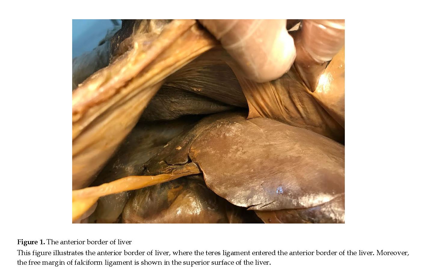
The general dissection course of abdominal wall of a 56-year-old man’s cadaver maintained in the North Khorasan University of Medicine in Iran, was investigated. We observed that a part of the liver falciform ligament was not formed and the liver had abnormal attachments to the anterior abdomen wall (Figure 1). Our research about the agenesis or defects of falciform ligament in the liver suggested that it is a rare anatomical variation, only described in limited studies. This report demonstrated a congenital defect of the falciform ligament in the liver.
Although there are numerous anatomical variations in the abdominal cavity organs, those associated with the liver ligament attachments are clinically important. One of which is a falciform ligament, that runs from the anterior abdominal wall to the diaphragm and attaches the liver to the umbilical ring [1]. Previous studies have suggested that anatomical variations in the thickness, stretch, and development of falciform ligament are different in adults and children [2]. According to previous research, the length and mean thickness of falciform ligament in adults are about 9.9 cm and 6 mm, respectively. Furthermore, this ligament encloses the obliterated umbilical vein [3].
A record of the liver ligaments anatomical variations is important for predicting laparoscopic abdominal surgery complications. Additionally, anatomical variations in the liver attachments and the right hypochondriac region can sometimes cause acute abdomen symptoms and medical emergency conditions like bowel obstruction [3, 4]. For example, sometimes a part of the bowel herniated through a defect in the falciform ligament may be caused by trocar insertion in operation room; also, part of small bowel herniated through a congenital defect in the falciform ligament may be created in a virgin abdomen [5].
However, data on the partial agenesis of falciform ligament are scarce. In this paper, we presented a case of anatomical congenital partial agenesis of this ligament in a 56-year-old formalin-fixed human body.
2. Case Presentation
The general dissection course of abdominal wall of a 56-year-old man’s cadaver maintained in the North Khorasan University of Medicine in Iran, was investigated. We observed that a part of the liver falciform ligament was not formed and the liver had abnormal attachments to the anterior abdomen wall (Figure 1). Our research about the agenesis or defects of falciform ligament in the liver suggested that it is a rare anatomical variation, only described in limited studies. This report demonstrated a congenital defect of the falciform ligament in the liver.
In other words, we observed that a part of the falciform ligament was not formed. However, the round ligament of liver was hung from the anterior abdominal wall above the umbilical ring and was inserted inside the parenchyma of the anterior border of liver in the normal position. Inspecting the diaphragmatic and visceral surface of liver revealed no hypertrophy or abnormal findings in the liver lobes. In addition, examining the abdominal wall and abdominal contents suggested no signs of surgical injury. Defects of falciform ligaments are important in surgery and sometimes in a part of the bowel herniated through this defect in the falciform ligament.
3. Discussion
Partial agenesis of the falciform ligament are extreme anatomical variations; this abnormality is important in clinical conditions. Defects generated in the falciform ligament may be congenital or the result of an abdominal surgical trauma. Sometimes, the surgical defects of falciform ligament present as a cystic abdominal mass or abscess [6]. Most reports of the abnormality, hypoplasia or failure of falciform ligament have been congenitally-related; no signs of abdominal surgery was observed in the current report [7].
Internal herniation through a congenital defect in this ligament may be caused by factors like pregnancy trauma that moves the abdominal contents into the upper abdomen and falciform ligament. We investigated the cadaver of a 56-year-old man with no signs of surgical incision to the abdominal wall. It seemed the huge defect in the falciform ligament was congenital. The variations in the falciform ligament anatomy are well-defined; however, disorders related to the falciform ligament remain undiscovered. Prevalent complications such as ligament cysts, tumors, abnormal vascularization, and partial ligament defects are the most recognized congenital abnormalities in the falciform ligament [8].
4. Conclusion
The diagnosed defects in the falciform ligament may indicate the importance of protective procedures to inhibit internal herniation through the falciform ligament.
Ethical Considerations
Compliance with ethical guidelines
This study was approved by the Ethics Committee of North Khorasan University of Medical Sciences (code: IR.NKUMS.REC.1398.002).
Funding
This research did not receive any specific grant from funding agencies in the public, commercial, or not-for-profit sectors.
Conflict of interest
The authors declared no conflict of interest.
Acknowledgements
I wish to thank my colleague, Dr Shahriar Ahmadpour for his assistance on taking pictures and performing cadaver dissection, as well as providing extremely useful comments.
References
Ozkececı ZT, Ozsoy M, Celep B, Bal A, Polat C. A rare cause of acute abdomen: An isolated Falciform ligament necrosis. Case reports in emergency medicine. 2014; 2014(570751):1-3. [DOI:10.1155/2014/570751]
Han SY. Variation in Falciform ligament with pheumoperitoneum. Journal of the Canadian Association of Radiologists. 1980; 31(3):171-3. [PMID]
Lakdawala M, Chaube SR, Kazi Y, Bhasker A, Kanchwala A. Internal hernia through an iatrogenic defect in the Falciform ligament: A case report. Hernia. 2009; 13(2):217-9. [DOI:10.1007/s10029-008-0424-7] [PMID]
Cagaš J, Vlček P, Jeřábek J. Rare internal hernia in the falciform ligament as a rare course of abdominal emergency and infrequent cause of ileus. Rozhledy v Chirurgii: Mesicnik Ceskoslovenske Chirurgicke Spolecnosti. 2012; 91(10):558-60. [PMID]
Elkhoury MI, El-Khoury F, Salloum E, Chahine EG, Chebib JE, Elhajj I, et al. Internal hernia as a complication of congenital falciform ligament window. Acta Chirurgica Belgica. 2013; 113(3):233-7. [DOI:10.1080/00015458.2013.11680919] [PMID]
Brock JS, Pachter HL, Schreiber J, Hofstetter SR. Surgical diseases of the Falciform ligament. The American Journal of Gastroenterology. 1992; 87(6):757-8. [PMID]
Dusu K, Dindyal S, Gadhvi V. Small bowel obstruction via herniation through an iatrogenic defect of the falciform ligament following laparoscopic cholecystectomy. Annals of the Royal College of Surgeons of England. 2015; 97(6):e93-5. [DOI:10.1308/rcsann.2015.0022] [PMID] [PMCID]
Macina S, Testa T, Losacco C. Congenital internal hernia through defect in the falciform ligament in adult: A case report and review of the literature. International Journal of Surgery Case Reports. 2016; 26:104-7. [DOI:10.1016/j.ijscr.2016.05.003] [PMCID] [PMID]
Type of Study: News and Reports |
Subject:
Gross Anatomy
Received: 2018/07/21 | Accepted: 2019/11/12 | Published: 2019/07/1
Received: 2018/07/21 | Accepted: 2019/11/12 | Published: 2019/07/1
Send email to the article author
| Rights and permissions | |
 |
This work is licensed under a Creative Commons Attribution-NonCommercial 4.0 International License. |
Contact Information
Anatomical Sciences Journal (ASJ)
Negah Institute for Scientific Communication, No.15, Na'eemi St., Mirzaye Shirazi St., Tehran, Iran.
Publisher Tel : +9821 4535 5555;
+9821 4535 5000
Website: http://www.anatomyjournal.ir/
E-mail: anatomyjournal@gmail.com
