Sat, Apr 20, 2024
Volume 19, Issue 1 (Winter & Spring 2022)
ASJ 2022, 19(1): 11-16 |
Back to browse issues page

Download citation:
BibTeX | RIS | EndNote | Medlars | ProCite | Reference Manager | RefWorks
Send citation to:


BibTeX | RIS | EndNote | Medlars | ProCite | Reference Manager | RefWorks
Send citation to:
Goudarzi A S, Ghorbani M, Esmaeili Gouvarchin Ghaleh H, Jalali Kondori B. Histopathological Assessment of Subacute Doses of Tebuconazole on Hepatotoxicity in Rats. ASJ 2022; 19 (1) :11-16
URL: http://anatomyjournal.ir/article-1-562-en.html
URL: http://anatomyjournal.ir/article-1-562-en.html
Amir Saeed Goudarzi1 

 , Masoud Ghorbani2 , Hadi Esmaeili Gouvarchin Ghaleh3 , Bahman Jalali Kondori 1
, Masoud Ghorbani2 , Hadi Esmaeili Gouvarchin Ghaleh3 , Bahman Jalali Kondori 1
1- Department of Anatomical Sciences, Faculty of Medicine, Baqiyatallah University of Medical Sciences, Tehran, Iran.
2- Applied Biotechnology Research Center, Research Institute of Biological Sciences and Non-agent Defense, Baqiyatallah University of Medical Sciences, Tehran, Iran.
3- Applied Virology Research Center, Research Institute of Biological Sciences and Non-agent Defense, Baqiyatallah University of Medical Sciences, Tehran, Iran.
Full-Text [PDF 1224 kb]
(367 Downloads)
| Abstract (HTML) (1189 Views)
Full-Text: (233 Views)
1. Introduction
Tebuconazole (TEB) is a systemic triazole fungicide that inhibits ergosterol biosynthesis in the cell membrane of fungi and stops the development of these pathogens [1]. TEB is today widely used in crops and ornamental plants. The use of pesticides in agricultural environments is a requirement for crop protection. On the other hand, the same safety for crops also endangers human and wildlife health [2]. TEB toxicity occurs when humans or animals have either direct exposure to the toxin or indirect exposure to infected plants. Symptoms of TEB toxicity have previously been described in rats, including restlessness, imbalance, spasm in some organs, and severe asthenia [3]. Metabolism of conazole-triazole fungicides occurs in hepatic microsomes [4].
Triazole fungicides increase the production of free radicals and oxidative stress in the body by significantly reducing glutathione-S transferase (GST) activity. Free radicals produced by oxidative stress lead to lipids, proteins, and DNA oxidation. These alterations eventually lead to cell death through apoptosis or necrosis, causing significant disorders in the body [5].
Moser et al. investigated the effect of TEB on rats’ immune, reproductive, and nervous systems in a study, according to which the impact on the nervous system was observed in the behavioral and neuropathological alterations [6]. Sancho et al. showed that short-term exposure to TEB at subacute doses causes physiological disorders in male fish, and the fungicide is a potential neurotoxin [7]. Recently, several case reports have been provided for the extensive hepatotoxic effects of TEB in animals and humans [8, 9]. Accordingly, it seems required to investigate the hepatotoxic effects of unintentional exposure to TEB. The results of this study point out the dangers of this drug and the need for more precautions when using it in agriculture.
2. Materials and Methods
Animals
Sixty male Wistar rats weighing 200-220 g were provided from the animal house of Baqiyatallah University of Medical Sciences, Tehran, Iran. The animals were kept under controlled conditions, including 22±2°C and a 12-12h light-dark cycle photoperiod. All institutional and national guidelines for the care and use of laboratory animals were followed. All experiments were performed following the principles of working with laboratory animals approved by the Ethics Committee of Baqiyatallah University of Medical Sciences.
Study design
The experimental rats were randomly divided into 6 groups (n=10 per group) using a random number table. Rats in each group were placed in special and similar cages of 40×20×20cm. One group was a control group, and the other 5 were considered experimental groups. TEB was administered for each experimental group, dosing by oral gavage at 6, 12, 25, 50, and 100 mg/kg for 30 days. To assess the short-term effects of the drug, five rats were randomly selected at the end of the first week from all groups and deeply anesthetized by injection of ketamine (100 mg/kg) and xylazine (10 mg/kg). A liver biopsy was then performed after blood sampling from the heart. Liver tissue samples were taken from other animals by the above method after the last dose of TEB at the end of the fourth week.
Biochemical analysis
The animals were anesthetized with ketamine-xylazine administration, and a direct blood sample was taken from the heart using a 5ml syringe, with 3-5 ml of blood from each animal. The blood samples were incubated at room temperature for a few minutes and then centrifuged at 5000 rpm for 10 min. Serum was separated from the clot, and samples were stored at -20°C to measure enzymes and biochemical parameters. Serum concentrations of AST, ALT, and ALP were measured by spectrophotometer, and the values of desired enzymes were measured by Pars Azmun ELISA kit.
Histopathological analysis
Liver samples were taken from all groups, and they were immediately placed in 10% formalin for fixation and preparation of tissue sections after rinsing with physiological serum. Tissue processing of fixed samples was performed, and tissue sections with a thickness of 5 μm were prepared as a serial section using a rotary microtome. Liver tissues in all study groups were stained with hematoxylin and eosin to analyze their histopathological alterations. Alterations in parameters such as cell inflammation, lipid vacuole accumulation, cell necrosis, disintegration in the portal space, and a considerable distance between cells were analyzed.
Statistic analysis
Data were analyzed by SPSS software using analytical statistics and one-way analysis of variance (One-way ANOVA) using a blind analysis method. The significant level was considered at P<0.05.
3. Results
Statistical analysis of the data showed that serum levels of AST, ALT, and ALP enzymes elevated in all doses of TEB compared to the control group; however, this elevation was not significant at all doses (Table 1).
.jpg)
The increase in ALT in the 50 and 100 mg/kg groups was statistically significant compared to the control group. The results also showed that the increase in AST and ALP was significant in the groups receiving 25, 50, and 100 mg/kg TEB compared to the control group.
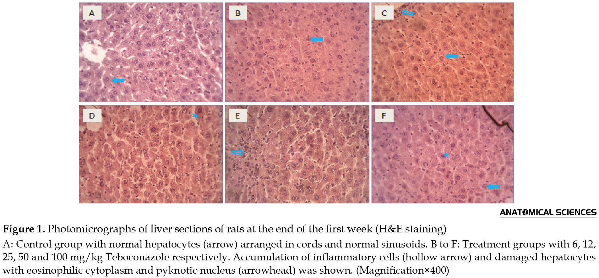
The histopathological analysis of the liver in different study groups indicated that the liver tissue structure was undamaged and normal in the control group. In the groups receiving oral TEB, various tissue alterations were observed, including accumulation of inflammatory cells around the central vein, sinusoidal dilatation, irregular hepatocyte plates (Remark string), and pyknotic hepatocyte in liver tissue, which were dose-dependent.
Assessment of the short-term (1 week) effects of TEB on liver tissue shows the hepatotoxic effects of this substance even at low doses (Figure 1).
Histopathological alterations of the liver in the long-term exposure (4 weeks) to TEB are shown in Figure 2.

The results suggested an increase in hepatocyte necrosis, destruction, and accumulation of lipid vacuoles and microvesicular steatosis in the study groups compared to short-term alterations.
4. Discussion
The current study showed that very low doses of TEB could cause significant hepatotoxic alterations in rats. According to our results, TEB elevates serum levels of AST, ALP, and ALT in rats. The elevation in these enzymes, indicators of liver damage, is directly related to the dose of TEB. The results of our study also showed various histopathological alterations in the liver tissue of animals exposed to TEB. The hepatotoxic effects of TEB in the early days are primarily seen in the accumulation of inflammatory cells and alterations in the order of hepatocytes and sinusoids. In the long-term exposure, these alterations were observed in the form of lipid accumulation, vascular and sinusoidal dilatation, and finally, extensive necrosis of hepatocytes.
As a result, the most destructive effects of these toxins are observed in the liver due to the metabolism location of conazole-triazole fungicides in liver microsomes. However, previous studies have also shown the destructive effects of TEB on other organs in the body. Ronis et al. in a study, identified multiple lesions of renal tissue exposed to TEB. This report indicates the occurrence of diffuse necrosis and severe hyperemia, which may be due to the effect of TEB on cells and their destruction through the mechanisms of inhibition and induction of cytochrome P450 [10]. Yargholi et al. investigated the histopathological effects of TEB on dog liver [8]. The researchers reported that the hepatocytes underwent adipose tissue changes, necrosis, and extensive destruction, which is consistent with our results.
The destructive effects of conazole-triazole fungicides have been reported to a limited extent in humans. Alaa et al. reported that oxidative stress and increased lipid peroxidation were observed in farm workers spraying conazole fungicide. This study showed that exposure to fungicides significantly reduces the activity of the glutathione-S transferase enzyme and induces oxidative stress in the human body by producing free radicals [11].
The hepatotoxic effects of TEB at subacute doses in humans were reported in another case report. In this case report, unintentional exposure of a farmer to a subacute dose of TEB caused a severe elevation in liver enzymes AST, ALP, and ALT in this person. The researchers concluded that unintentional exposure to low doses of TEB could increase an individual’s risk of developing hepatitis [9]. Our study results also showed a sharp elevation in liver enzymes during exposure to TEB, which was consistent with this study.
5. Conclusion
The results of our study showed significant hepatotoxic effects at subacute doses of tebuconazole. It is recommended to take the required precautions, including personal protection tools, due to the hazardous effects of this fungicide. It is also recommended to use other fungicides with less toxicity to mammals.
Ethical Considerations
Compliance with ethical guidelines
The Ethics Committee of Baqiyatallah University of Medical Sciences approved this study (Ethical ID: IRIR.BMSU.REC.1398.384).
Funding
This research was supported by the research project (Grant No.: 98000386) of the Baqiyatallah University of Medical Sciences.
Authors' contributions
All authors equally contributed to preparing this article.
Conflict of interest
The authors declared no conflict of interest.
Acknowledgments
We would like to thank the Student Research Committee of Baqiyatallah University of Medical Sciences, for financial support.
References
Tebuconazole (TEB) is a systemic triazole fungicide that inhibits ergosterol biosynthesis in the cell membrane of fungi and stops the development of these pathogens [1]. TEB is today widely used in crops and ornamental plants. The use of pesticides in agricultural environments is a requirement for crop protection. On the other hand, the same safety for crops also endangers human and wildlife health [2]. TEB toxicity occurs when humans or animals have either direct exposure to the toxin or indirect exposure to infected plants. Symptoms of TEB toxicity have previously been described in rats, including restlessness, imbalance, spasm in some organs, and severe asthenia [3]. Metabolism of conazole-triazole fungicides occurs in hepatic microsomes [4].
Triazole fungicides increase the production of free radicals and oxidative stress in the body by significantly reducing glutathione-S transferase (GST) activity. Free radicals produced by oxidative stress lead to lipids, proteins, and DNA oxidation. These alterations eventually lead to cell death through apoptosis or necrosis, causing significant disorders in the body [5].
Moser et al. investigated the effect of TEB on rats’ immune, reproductive, and nervous systems in a study, according to which the impact on the nervous system was observed in the behavioral and neuropathological alterations [6]. Sancho et al. showed that short-term exposure to TEB at subacute doses causes physiological disorders in male fish, and the fungicide is a potential neurotoxin [7]. Recently, several case reports have been provided for the extensive hepatotoxic effects of TEB in animals and humans [8, 9]. Accordingly, it seems required to investigate the hepatotoxic effects of unintentional exposure to TEB. The results of this study point out the dangers of this drug and the need for more precautions when using it in agriculture.
2. Materials and Methods
Animals
Sixty male Wistar rats weighing 200-220 g were provided from the animal house of Baqiyatallah University of Medical Sciences, Tehran, Iran. The animals were kept under controlled conditions, including 22±2°C and a 12-12h light-dark cycle photoperiod. All institutional and national guidelines for the care and use of laboratory animals were followed. All experiments were performed following the principles of working with laboratory animals approved by the Ethics Committee of Baqiyatallah University of Medical Sciences.
Study design
The experimental rats were randomly divided into 6 groups (n=10 per group) using a random number table. Rats in each group were placed in special and similar cages of 40×20×20cm. One group was a control group, and the other 5 were considered experimental groups. TEB was administered for each experimental group, dosing by oral gavage at 6, 12, 25, 50, and 100 mg/kg for 30 days. To assess the short-term effects of the drug, five rats were randomly selected at the end of the first week from all groups and deeply anesthetized by injection of ketamine (100 mg/kg) and xylazine (10 mg/kg). A liver biopsy was then performed after blood sampling from the heart. Liver tissue samples were taken from other animals by the above method after the last dose of TEB at the end of the fourth week.
Biochemical analysis
The animals were anesthetized with ketamine-xylazine administration, and a direct blood sample was taken from the heart using a 5ml syringe, with 3-5 ml of blood from each animal. The blood samples were incubated at room temperature for a few minutes and then centrifuged at 5000 rpm for 10 min. Serum was separated from the clot, and samples were stored at -20°C to measure enzymes and biochemical parameters. Serum concentrations of AST, ALT, and ALP were measured by spectrophotometer, and the values of desired enzymes were measured by Pars Azmun ELISA kit.
Histopathological analysis
Liver samples were taken from all groups, and they were immediately placed in 10% formalin for fixation and preparation of tissue sections after rinsing with physiological serum. Tissue processing of fixed samples was performed, and tissue sections with a thickness of 5 μm were prepared as a serial section using a rotary microtome. Liver tissues in all study groups were stained with hematoxylin and eosin to analyze their histopathological alterations. Alterations in parameters such as cell inflammation, lipid vacuole accumulation, cell necrosis, disintegration in the portal space, and a considerable distance between cells were analyzed.
Statistic analysis
Data were analyzed by SPSS software using analytical statistics and one-way analysis of variance (One-way ANOVA) using a blind analysis method. The significant level was considered at P<0.05.
3. Results
Statistical analysis of the data showed that serum levels of AST, ALT, and ALP enzymes elevated in all doses of TEB compared to the control group; however, this elevation was not significant at all doses (Table 1).
The increase in ALT in the 50 and 100 mg/kg groups was statistically significant compared to the control group. The results also showed that the increase in AST and ALP was significant in the groups receiving 25, 50, and 100 mg/kg TEB compared to the control group.
The histopathological analysis of the liver in different study groups indicated that the liver tissue structure was undamaged and normal in the control group. In the groups receiving oral TEB, various tissue alterations were observed, including accumulation of inflammatory cells around the central vein, sinusoidal dilatation, irregular hepatocyte plates (Remark string), and pyknotic hepatocyte in liver tissue, which were dose-dependent.
Assessment of the short-term (1 week) effects of TEB on liver tissue shows the hepatotoxic effects of this substance even at low doses (Figure 1).
Histopathological alterations of the liver in the long-term exposure (4 weeks) to TEB are shown in Figure 2.
The results suggested an increase in hepatocyte necrosis, destruction, and accumulation of lipid vacuoles and microvesicular steatosis in the study groups compared to short-term alterations.
4. Discussion
The current study showed that very low doses of TEB could cause significant hepatotoxic alterations in rats. According to our results, TEB elevates serum levels of AST, ALP, and ALT in rats. The elevation in these enzymes, indicators of liver damage, is directly related to the dose of TEB. The results of our study also showed various histopathological alterations in the liver tissue of animals exposed to TEB. The hepatotoxic effects of TEB in the early days are primarily seen in the accumulation of inflammatory cells and alterations in the order of hepatocytes and sinusoids. In the long-term exposure, these alterations were observed in the form of lipid accumulation, vascular and sinusoidal dilatation, and finally, extensive necrosis of hepatocytes.
As a result, the most destructive effects of these toxins are observed in the liver due to the metabolism location of conazole-triazole fungicides in liver microsomes. However, previous studies have also shown the destructive effects of TEB on other organs in the body. Ronis et al. in a study, identified multiple lesions of renal tissue exposed to TEB. This report indicates the occurrence of diffuse necrosis and severe hyperemia, which may be due to the effect of TEB on cells and their destruction through the mechanisms of inhibition and induction of cytochrome P450 [10]. Yargholi et al. investigated the histopathological effects of TEB on dog liver [8]. The researchers reported that the hepatocytes underwent adipose tissue changes, necrosis, and extensive destruction, which is consistent with our results.
The destructive effects of conazole-triazole fungicides have been reported to a limited extent in humans. Alaa et al. reported that oxidative stress and increased lipid peroxidation were observed in farm workers spraying conazole fungicide. This study showed that exposure to fungicides significantly reduces the activity of the glutathione-S transferase enzyme and induces oxidative stress in the human body by producing free radicals [11].
The hepatotoxic effects of TEB at subacute doses in humans were reported in another case report. In this case report, unintentional exposure of a farmer to a subacute dose of TEB caused a severe elevation in liver enzymes AST, ALP, and ALT in this person. The researchers concluded that unintentional exposure to low doses of TEB could increase an individual’s risk of developing hepatitis [9]. Our study results also showed a sharp elevation in liver enzymes during exposure to TEB, which was consistent with this study.
5. Conclusion
The results of our study showed significant hepatotoxic effects at subacute doses of tebuconazole. It is recommended to take the required precautions, including personal protection tools, due to the hazardous effects of this fungicide. It is also recommended to use other fungicides with less toxicity to mammals.
Ethical Considerations
Compliance with ethical guidelines
The Ethics Committee of Baqiyatallah University of Medical Sciences approved this study (Ethical ID: IRIR.BMSU.REC.1398.384).
Funding
This research was supported by the research project (Grant No.: 98000386) of the Baqiyatallah University of Medical Sciences.
Authors' contributions
All authors equally contributed to preparing this article.
Conflict of interest
The authors declared no conflict of interest.
Acknowledgments
We would like to thank the Student Research Committee of Baqiyatallah University of Medical Sciences, for financial support.
References
- Shen Z, Zhu W, Liu D, Xu X, Zhang P, Zhou Z. Stereoselective degradation of tebuconazole in rat liver microsomes. Chirality. 2012; 24(1):67-71. [DOI:10.1002/chir.21027] [PMID]
- Aktar MW, Sengupta D, Chowdhury A. Impact of pesticides use in agriculture: Their benefits and hazards. Interdisciplinary Toxicology. 2009; 2:1-12. [DOI:10.2478/v10102-009-0001-7] [PMID] [PMCID]
- Fustinoni S, Mercadante R, Polledri E, Rubino F, Mandic-Rajcevic S, Vianello G, et al. Biological monitoring of exposure to tebuconazole in winegrowers. Journal of Exposure Science & Environmental Epidemiology. 2014; 24(6):643-9. [DOI:10.1038/jes.2014.14] [PMID]
- Ward W, Delker D, Hester S, Thai s-f, Wolf D, Allen J, et al. Transcriptional Profiles in liver from mice treated with hepatotumorigenic and nonhepatotumorigenic triazole conazole fungicides: Propiconazole, triadimefon, and myclobutanil. Toxicologic Pathology. 2006; 34:863-78. [DOI:10.1080/01926230601047832] [PMID]
- Torres MCL, Soares NdFF, Maia JF. Kinetics parameters of Glutathione S-Transferase and its activation by vegetable extracts. Food Science and Technology. 2004; 24(2):243-8. [DOI:10.1590/S0101-20612004000200014]
- Barone S, Moser V. The effects of perinatal tebuconazole exposure on adult neurological, immunological, and reproductive function in rats. Toxicological Sciences. 2004; 77(1):183. [DOI:10.1093/toxsci/kfh036] [PMID]
- Sancho E, Villarroel M, Fernández C, Andreu-Moliner E, Ferrando M. Short-term exposure to sublethal tebuconazole induces physiological impairment in male zebrafish (Danio rerio). Ecotoxicology and Environmental Safety. 2009; 73(3):370-6. [DOI:10.1016/j.ecoenv.2009.09.020] [PMID]
- Yargholi M, Ali Esfehani T, Musavi Ss, Erfani AM, Salar Amoli J. [Poisoning of dog by tebuconazole fungicide-a case report (Persian)]. Iranian Veterinary Journal. 2016; 11(4):114-9. [DOI:10.22055/IVJ.2016.13022]
- Habibzadeh S, Yazdanbod A, Mohammadshahi J, Maleki N, Ataei S. Hepatotoxicity after exposure to tebuconazole: A case report and brief review.Hepatitis Monthly. 2019; 19(7):e94548. [DOI:10.5812/hepatmon.94548]
- Ronis MJJ, Celander M, Badger TM. Cytochrome P450 enzymes in the kidney of the bobwhite quail (Colinus virginianus): Induction and inhibition by ergosterol biosynthesis inhibiting. Comparative Biochemistry and Physiology Part C: Pharmacology, Toxicology, and Endocrinology. 1998; 121(1):221-9. [DOI:10.1016/S0742-8413(98)10043-9]
- Mecdad AA, Ahmed MH, ElHalwagy MEA, Afify MMM. A study on oxidative stress biomarkers and immunomodulatory effects of pesticides in pesticide-sprayers. Egyptian Journal of Forensic Sciences. 2011; 1(2):93-8. [DOI:10.1016/j.ejfs.2011.04.012]
Type of Study: Original |
Subject:
Histology
Received: 2022/01/10 | Accepted: 2022/02/9 | Published: 2022/01/1
Received: 2022/01/10 | Accepted: 2022/02/9 | Published: 2022/01/1
Send email to the article author
| Rights and permissions | |
 |
This work is licensed under a Creative Commons Attribution-NonCommercial 4.0 International License. |
Contact Information
Anatomical Sciences Journal (ASJ)
Negah Institute for Scientific Communication, No.15, Na'eemi St., Mirzaye Shirazi St., Tehran, Iran.
Publisher Tel : +9821 4535 5555;
+9821 4535 5000
Website: http://www.anatomyjournal.ir/
E-mail: anatomyjournal@gmail.com
