Tue, Apr 23, 2024
Volume 18, Issue 1 (Winter & Spring 2021)
ASJ 2021, 18(1): 31-44 |
Back to browse issues page

Download citation:
BibTeX | RIS | EndNote | Medlars | ProCite | Reference Manager | RefWorks
Send citation to:


BibTeX | RIS | EndNote | Medlars | ProCite | Reference Manager | RefWorks
Send citation to:
Rafiemanzelat F, Sheikhi M, Rabiei A A, Setayeshmehr M. P96 Technique for Tissue Plastination in Preparing Long-Lasting Anatomical Specimens. ASJ 2021; 18 (1) :31-44
URL: http://anatomyjournal.ir/article-1-245-en.html
URL: http://anatomyjournal.ir/article-1-245-en.html


1- Department of Chemistry, Organic Polymer Chemistry Research Laboratory, University of Isfahan, Isfahan, Iran.
2- Department of Anatomical Sciences, School of Medicine, Isfahan University of Medical Science, Isfahan, Iran.
2- Department of Anatomical Sciences, School of Medicine, Isfahan University of Medical Science, Isfahan, Iran.
Full-Text [PDF 3319 kb]
(1086 Downloads)
| Abstract (HTML) (1867 Views)
Full-Text: (1258 Views)
1. Introduction
For numerous centuries, anatomists have sought effective and safe techniques for the long-term preservation of biological tissues to prepare specimens suitable for teaching anatomical sciences and preserve the museum specimens [1]. Various techniques, including mummification, fixation, deparaffinization, embedding in epoxy-resin, Thiel embalming method, and plastination has been used for such purposes [1-3]. All these techniques are associated with advantages and disadvantages [2, 4-6]. For example, Thiel-embalmed cadavers have provided quality, elasticity, transparency, and handling efficiency, as life-like tissues. Thiel’s method possesses an exceptional sterilizing efficacy; however, it is limited up to 3 years and cadavers should be sealed in plastic bags or remain in submersion. The minority of experts in the method, relatively higher costs, and taking the fixation to step up to 6 months are the other disadvantages of Thiel’s method [2].
Plastination is a technique for preserving biological tissues, i.e., first introduced by Von Hagens in 1977. This technique provides dry, odorless, and durable specimens [7]. The plastinated specimens are resistant to mechanical damages resulting from passing through numerous hands, requiring minimal preservation [8]. There are some different plastination techniques to prepare various specimens suitable for altered purposes. These specimens demonstrated a widespread application in anatomy, pathology, neuroanatomy, veterinary sciences, radiology, zoology, biology, and forensic medicine [6, 9-13].
However, plastination is an expensive technique due to the necessary equipment and materials. Moreover, gaining or preparing materials, such as BIODUR S10, S6, S3, BIODURepoxy E12, E1, polyester P35, and P40 [6, 14-17] can be challenging for some countries. Thus, there were diverse efforts to introduce different materials and methods to achieve cost-effective techniques that can be attainable everywhere [4, 18-28]. For example, novel synthesized polyester resins (P87, P88, P89) which was developed in our group [21, 22], OR-TECH PR-10 silicone, i.e., locally formulated for the USA customers [25], Su-Yi Chinese silicone for plastination procedure at room temperature [26, 27], etc. were employed to achieve proper and available plastination techniques.
In this work, a novel Unsaturated Polyester Resin (UPR) was formulated. Furthermore, its quality was tuned to achieve the desired properties for organ plastination at Room Temperature (RT). This investigated method was intended to allow the main steps of conventional plastination techniques, including fixation, dehydration, forced impregnation, and curing, to be directed at RT using available materials and equipment. This study overcomes some disadvantages of synthesized polyesters, P87, P88, and P89, such as discoloration, yellowing, shrinkage, rigidity, and the obligation for the admixing of curing initiator after the impregnation step [21, 22]. The admixing of the curing initiator after the impregnation step resulted in the incomplete hardening process as well as sample tackiness even after the curing reaction. For further evaluation of the synthesized resin, some properties of P96 were compared with P35, as a commercially prepared resin.
2. Materials and Methods
Unsaturated Polyester Resins (UPR) were synthesized via the reaction of various polyols, consisting of Propyleneglycol (PG), Ethylene Glycol (EG), Diethylene Glycol (DEG), or Polyethylene Glycol (PEG) with different dibasic acids, such as Phthalic Anhydride (PhA), Adipic Acid (Ad), Sebacic Acid (Se), or Terephthalic Acid (TPh). Maleic Anhydride (MA), Tetrahydrophthalicanhydride (THPhA), or Fumaric Acid (FA) were used as sources of the unsaturated double bond. All the materials were commercially available grades and used as received. The general synthesis method was as follow:
The polymerization reaction underwent an esterification reaction between monomers. Briefly, MA1.49 mole (146.1gr), PhA1.49 mole (220.6gr), as well as PG and EG each 2.35 mole (178.7gr & 249.2gr, respectively) were poured into a three-necked 1litter flask. Then, 0.05×10⁻⁴ mole p-toluene sulfonic acid as catalyst and 1.13×10⁻³ mole hydroquinone as a radical scavenger were added into the reactor. The mixture was stirred (by a mechanical mixer, Heidolph, type 50111, Germany) at 105-110°C for 24 hours. A flux of nitrogen was passed through the solution. The resulting polymer was cooled under nitrogen to 85˚C at the end of the reaction. Then, 5.66×10⁻⁴ mole hydroquinone was added as an inhibitor to avoid the unintentional polymerization of the product. The temperature of the reaction mixture was decreased to 60˚C and Methyl Methacrylate (MMA) or Styrene (St) 30% wt (by the weight of the synthesized UPR) as an intermediate solvent, as well as a curing agent, was added dropwise to the mixture. P96-M or P96-S is the code assigning the prepared polymer samples with MMA or St, respectively.
To achieve proper formulation with desirable properties concerning the tissue color, contrast, the appearance of tissue texture, as well as suitable strength and flexibility, various raw materials, including glycols, diacids, and anhydrides were substituted in the above-mentioned procedure. Then, the interaction of the resulted polymers with various tissues was examined.
The crosslinking (hardening) reaction of the mold cast P96 resins were conducted at RT in a UV cabinet equipped with a UV-A lamp (400W, 365 nm, Noor Company, Iran). Photo curing reaction was conducted by the UV exposure of the samples at a 30cm distance from the lamp. The UV cabinet was equipped with two fans and an N2 purge inlet and outlet; they were placed at the bottom and top of the cabinet to circulate N2 gas and prevented temperature increase.
To determine the suitable curing agent, initiators, and promoters, P96-M or P96-S was mixed with different kinds and amounts of initiators and promoters. Then, the formulation was photo-cured after mold casting (Figure 1).
.PNG)
.PNG)
.PNG)
The formulation resulted in minimum tissue discoloration; high transparency; maximum gel content (complete curing reaction); proper flexibility; desirable toughness; suitable interaction with tissue to preserve its natural texture; color during impregnation, and hardening process was selected as the optimum formulation (Figure 1). Benzoyl peroxide, t-Butyl peroxide, Cobalt Octoate/Methyl Ethyl Ketone Peroxide (CO/MEKP), 2,2’-azobis (isobutyronitrile) (AIBN), acetophenone, benzophenone, benzil, 4,4’-Dichlorobenzophenone, Benzoin, and Camphorquinone (CQ) were among the examined initiators.
After determining the optimum formulation, the tissue specimens were impregnated using P96-S or P96-M. After the completion of impregnation, the tissue specimens were removed from the vacuum chamber and hung at RT to remove the excess resin (for bulk plastination). Then, the UV curing process of impregnated specimens was conducted according to the above-mentioned hardening procedure. The P87 samples, according to its formula, were cured in the presence of St as the curing agent and CO/MEKP as the curing initiator at 45˚C for 24h. The P35-impregnated samples were cured by the same procedure.
Different specimens obtained from sheep and rats were used as the study materials. The human organs were obtained from the donated cadavers at the Department of Anatomical Sciences, Faculty of Medicine (Isfahan, Iran). The specimens were processed via the conventional plastination method, consisting of fixation, dehydration (defatting if necessary), force impregnation, embedding, and hardening steps.
For the impregnation step, the specimens were immersed in a resin bath, containing a mixture of P96-M (or P96-S) and initiators, i.e., placed within the vacuum chamber. The pressure was reduced up to 10 mm Hg to force impregnate the tissues by resin via negative pressure, i.e., resulted from the vaporization of the acetone. An intermittent vacuum procedure was used which resulted in declined capital costs required for a high-quality vacuum pump and expensive spark-proof deep freezer. After the complete replacement of acetone by resin (about 1-14 days depending on the size of the specimens), the specimens were taken out of the vacuum chamber. The specimens were hung to drain back the excess resin into the chamber (for bulk plastination).
For the sheet plastination protocol, the specimens, after fixation, were submerged in tap water for 2h to remove fixative materials. Then, they were carefully cut sagittal, coronal, or horizontally using a special band saw at -196°C Finally, the specimens were cast via the sandwich method following force impregnation.
The flexibility of the cured resin, its stiffness, and tensile strength was determined using an instrument (SANTAM-SMT5 model) according to ASTM D412-C. To measure the relevant mechanical properties (tensile test), the samples (P96, P87, or P35 resins) were cured in a mold with the dimensions of 115mm total length, 25mm total width, 33mm gage length, 6mm gage width, and 3mm thickness (Figures 2 & 3). The tensile strength (stress) was calculated by dividing the maximum load in Newton (N) by the average original cross-sectional area of the specimens in square meters. The elongation percent (strain %) was calculated by dividing the variation in the sample gage length (Δl) to the original specimen gage length (l0), expressed as a percentage (%). For these tests, 24 pieces prepared in the same block were used (n=8/group, G1=P87, G2=P35, G3=P96). In the tensile test, for comparing the samples’ strength, we determined the maximum force to break the specimen at constant speed (V=50 mm/min) by the extensometer.
For the weight test, 24 slices (n=8/group), after mold curing, were cut into the same block size, 3×1×1cm, and weighed. The density was obtained by dividing the sample weight by its volume.
To measure the shelf-life of different formulations, the vessel, containing complete formulation, including polyester, curing agent, initiator, and promoter (co-initiator if necessary) was put at the temperature of 50˚C as well as at RT. Besides, the necessary time passed for the occurrence of any possible changes in the physical state of the sample (increasing viscosity, gelation phenomena) was measured.
The gel content of the cured polymeric sheets was determined by computing the weight loss after 24h extraction with acetone at reflux condition, according to the ASTM D2765-84 technical standard. The weight loss (W0-W) was considered as the portion of the non-crosslinked polymer, which can be related to the incomplete hardening process. Accordingly, a cured polymeric sheet with a definite weight was rinsed in acetone for 24h under a Soxhlet extractor, and the final Weight (W) was measured after drying. Measuring gel content can be performed by Equation 1: where the W0 is the initial weight and W is the final weight after solvent extraction.
1. Gel content % = [W/W0] × 100 eq
The viscosity (cp) of the synthesized resin was measured using a Brookfield viscometer (model DVIII) equipped with a spindle-34 and a thermostat at 25°C.
Texture Analyzer TA.XT (Stable Micro Systems Ltd., Surrey, UK) was used to determine the cohesiveness (consistency) and resilience (flexibility) properties of the plastinated specimens. A 25mm (diameter) disk of the sample was compressed by the rate of 4 mm/s and texture properties were evaluated according to a TPA formula.
The linear shrinkage of the synthesized resin was determined according to the ASTM D2566-86 standard. The linear shrinkage (l) was changed to the volume percentage (v) according to Equation 2. The obtained results were reported as the mean values of 8 repeated specimens, i.e., simultaneously cured. Linear shrinkage was measured via casting the resin into the Teflon troughs of the semicircular cross-section with fiat ends. Cured resin specimens were removed from the troughs after cooling to room temperature. Moreover, their lengths were determined using a caliper gauge. Shrinkage Δl/l0 was calculated as the length-wise difference between the trough and cured sample at 23°C, divided by the length of the trough l0.
2. ΔV/V0 = [(1+Δl/l0)3 – 1]× 100 eq
LAB colorimetric index was employed to report the resulted color after the crosslinking of the resin and the plastinated samples. Accordingly, a colorimetric device equipped with two fluorescent lamps and a camera (Panasonic, Lumix DMC-TZ5) was applied.
The odor and quality of the t plastinated specimens produced via P87 and P96 techniques were evaluated using a checklist completed by anatomy experts and students; they were requested to rate the odor and quality of the specimens for educational and research purposes on a scale of 0-10. In this scale, 0-1=weak, 2-4=medium, 5-7=high, and 8-10=utmost as per the criteria, i.e., under evaluation.
A) Odor: The plastinated specimens are odor.
B) Educational quality: The specimen has maintained its natural texture and color; it is durable and it is not shrinkage and sticky.
The collected results were analyzed using SPSS. Differences with P<0.05 were interpreted as significant. To compare the results of tensile and density tests between each pair (P96/P35) and (P96/P87) among the 3 groups (n=8) of P96, P87, P35 of plastinated specimens or molded cured resins, a t-test was used. Wilcoxon signed-rank test was employed to compare the results of the odor, educational quality, and stickiness, between P96 and P87 of plastinated specimens. The two groups (n=8) of plastinated specimens consisted of heart plastinated with P87 or P96 resin were considered.
3. Results
The mean values of tensile stress (force ÷ cross-area), elongation (strain (Δl ÷ l0)), tensile module (stiffness), energy (toughness), and bending data resulted from the tensile test as well as density data resulted from weight test are presented in Table 1.
.PNG)
The results of the t-test and Levene’s test are presented in Table 2. Table 2 lists the level of significance to compare each pair of samples per study variable.
The results of the weight test indicated that the P96 samples were lighter, compared to the P35 and P87 ones. The weight of the cured blocks prepared with P96 was about one-half of the weight of P35 samples (Table 1). There was a significant difference (P≤0.05) between the mean densities of samples prepared via P96, compared to P35. There was no significant difference between the Mean±SD density of P87(1.1647±0.08) and P96 samples (1.1625±0.05) (P>0.05).
The tensile test data indicated that P96 samples presented higher elongation, compared to P87and P35 samples; however, the required force for its strain was lower than that of P87 and P35 samples. This can be confirmed by comparing their stress and module data. The required bending force of P96 samples was also lower than that of P87and P35 samples. These data supported the greater flexibility of P96 samples, compared to P87 and P35 samples for bulk plastination.
The collected data revealed that the differences in the mean values of all study variables were significant between P96 and P35 (P≤0.05). Furthermore, the mean differences of all study variables were significant between P96 and P87 (P≤0.05), except for density (P=0.950).
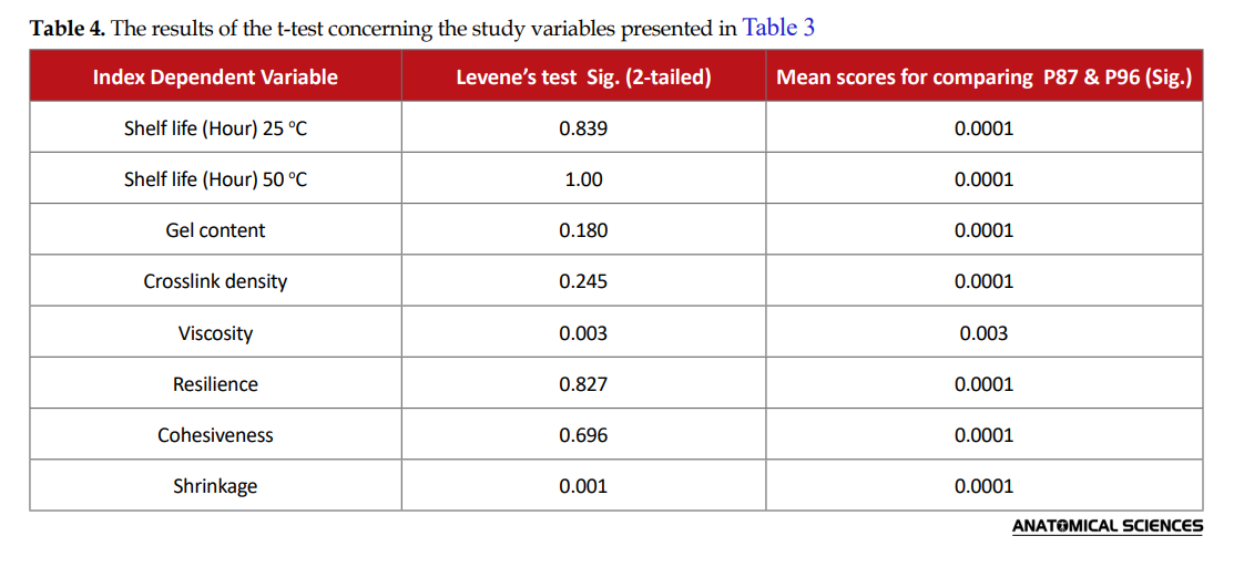
The mean levels of shelf life, gel content, crosslinking density, viscosity, resilience, cohesiveness, and shrinkage values recorded for two groups of materials (P87 and P96) are presented in Table 3.
.PNG)
The shelf life of different formulations of resins, including polyester, curing agent, initiator, and promoter, reflects the possibility of storing the specific formulation after synthesis and their storage conditions. The relevant data signified no further possibility for the storage of the complete formulation of P87 at RT. This is because the gelation and hardening process occurs soon after adding initiator and promoter to the polyester resin containing curing agent. In other words, during embedding and impregnation steps at RT, initiator and promoter cannot be uniformly mixed with P87 formulation in impregnation bath. Thus, only after the impregnation step, initiator and promoter can be applied to the specimen surfaces by a brush. This can result in the non-uniform mixing of reactive components into the depth of the impregnated tissue and an incomplete hardening process, especially for thick samples. New synthesized formulation P96, has presented storage possibility for a long time without any special storage conditions. However, this facilitates the uniform mixing of all reactive components during the impregnation step. Furthermore, curing will only initiate after UV exposure.
Gel content and crosslink density can be related to the degree of the completion of the hardening process. The more complete the process is, the fewer unreacted components will remain in the prepared samples. The presence of unreacted components will result in the sticky specimen’s surface, opaque product, the leakage of the unreacted materials to the specimen’s surface, and reduce the mechanical strength of the final product. According to the gel content values, after one month of the hardening process, the P96 materials revealed a more complete hardening process, compared to P87 materials (Table 3). Additionally, higher gel content makes the polymer more resistant against water and fungi and increases its durability.
The viscosity of formulated resins indicates the ease of penetration of the resin into the tissue during the impregnation step. The lower viscosity resulted in the easier and faster penetration of the resin into the tissue. The achieved results reflected that the viscosity of the P87 formulation was faintly lower than that of P96. According to the texture analysis data, the resilience values obtained for two groups of the P87 and P96 plastinated heart samples indicated that the P96 samples were more elastic than the P87 ones. The P87 specimens were approximately 3.5 times stickier than P96 specimens per comparing their cohesiveness (stickiness) values (Table 4).
The collected results suggested that the shrinkage of the cured P96 samples was <4%, highlighting the very immaterial deformation of the specimen.
LAB colorimetric index was used to report the resulted color of the samples after crosslinking. The LAB color space was modeled after a color-opponent theory stating that two colors cannot concurrently be red and green or yellow and blue. L indicates lightness, a is the red/green coordinate, and b is the yellow/blue coordinate. Deltas for l(ΔL), a (Δa), and b (Δb) may be positive (+) or negative (-). The total difference, Delta E (ΔE), however, always remains positive.
Δl (l sample minus lstandard) = difference in lightness and darkness (+ = lighter, - = darker)
Δa (a sample minus a standard) = difference in redness and greenness (+ = redder, - = greener)
Δb (b sample minus b standard) = difference in yellow and blue (+ = yellower, - = bluer)
ΔE = total color difference = [ΔL2 + Δa2 + Δb2]1/2
Comparing P87 (as standard) to P96 (as sample) upon l, a, and b values per product (Table 5), we can objectively determine that the two products were not matched in color. This conclusion was supported by data analysis signifying that the difference of mean values of l, a, and b data was significant between P96 and P87 (P≤0.05) (Table 6). The heart specimens plastinated by P96 suggested more lightness, redness, and yellowness compared to specimens plastinated by P87 (Table 5, Figure 4).
.PNG)


The results of the whole body plastination demonstrated that darkness, greenness, and blueness values obtained for P87 are more obvious than those of P96 (Figure 5).

The quality and odor of the P96 and P87 plastinated specimens were evaluated via the data extracted from the checklist. P96 plastinated specimens (group I) were compared to the corresponding P87 specimens (group II). The responses were recorded as weak, medium, high, or utmost per the criteria under evaluation. The specimens used in this experiment were evaluated one year after plastination. This is because P87 fresh plastinated samples were very odor and sticky.
The Wilcoxon signed-rank test was used to compare the results of the checklist regarding the quality of P96 and P87 samples. The related results presented a significant difference between the qualities of the specimens plastinated via the two plastination methods (P=0.025). The results suggested that the P96 plastination method effectively improved the quality level. However, there was no significant difference between the odor of the specimens plastinated via P96 and P87 methods (P=0.096) (Table 7).

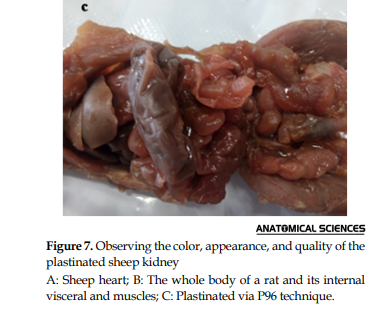
Following the impregnation, after the hardening process, all specimens were sticky and odor. However, after completing the hardening process, depending on the type of the resins and thickness of the samples, the resulted specimens were dry, relatively odorless, tough, and durable (Figures 4, 5, 6 & 7).
According to the data extracted from the checklists, the visual comparison of the samples indicated that bulk specimens plastinated via the P96 technique preserved their natural texture and color. Furthermore, they were flexible as well. These specimens, immediately after the completion of the curing process, were touchable, non-sticky, and dry. The P87 samples were tough and touchable; however, even one year after curing, they were still slightly sticky. Moreover, after one-month post-hardening step, the P96 samples were more odorless, compared to the P87 samples.
As shown in Figure 6, the detailed anatomical structure of the brain, like the brush appearance of corona radiate is observed on the brain slides plastinated via P87 and P96 methods. The P87 and P96 plastinated slices presented an excellent distinction between white and gray matter after one year. Additionally, the P96 sheet was clearer than the P87 or P35 sheet.

The P96 specimens were stored in the anatomy museum for more than 2 years while the temperature varied between 15-36°C. However, there were no signs of fungal infection in the P96 samples, based on the lack of change in the color and smell of the samples (Figures 4, 5, 6 & 7).
4. Discussion
Understanding the structure and variation of the anatomic organ is essential for nurses, physicians as well as specialists, such as surgeons or radiologists [29]. Plastination is a modern technique in which polymers are replaced with tissue water to preserve tissues close to the original form [6, 30]. Gunther von Hagens presented three classic forms of plastination techniques named in connection with the using materials; S10, P40, and E12 [31]. Plastinated organs are widely used to teach gross anatomy in medical and veterinary colleges. Plastination as a teaching method of animal science can cause fewer animals to be sacrificed. Plastination produces long-lasting, easily touchable, non-hazardous, and almost natural-looking tissue specimens [7]. S10 technique is mostly used for the bulk plastination of internal organs and joint preparations [30].
The high cost of silicone resin, the need for low-temperature equipment, and a gas chamber for the hardening process following the increases in demand for plastinated specimens have encouraged researchers for using low-cost alternative materials and methods for the long-term preservation of the biological tissues. Other common disadvantages of the S10 technique are shrinkage, the change of color, visible defects, which include the deformation, damaged arteries, and nerves, or rough surface of specimen [7]. P35 and P40 plastination techniques are mostly used for making plates of the brain with a thickness of 0.3-20mm. These techniques require no pre-staining of brain tissue. This is because it causes a strong contrast between the gray and white matter of the cerebrum. P35 and P40 polyesters techniques are specific approaches to brain sheet plastination and have limited usage for bulk plastination [31, 32]. They ultimately include some long preparative stage of the head and fixing in several fluids [32]. E12 epoxy plastination technique is suitable for making topographic-anatomical slices with the thicknesses of 0.7-1.2mm of the limbs and trunk. The resulting sheets are transparent and resistant to mechanical stress; however, they are hard, brittle, and nonflexible [31]. Furthermore, epoxy resin sheets tend to turn yellow over several years [32]. Thus, it needs to be placed in an oven at 45°C for 4 days for hardening. Additionally, it requires numerous stages of fat removal from the tissue to gain the best optical quality in the plastinated sections [33]. Ravikumar examined a new method of sheet plastination which was mainly applied for preserving the tissue slices of a dolphin using a polyester resin. Towards the end of the method, the cast molded slices were cured in a heated water bath [32].Gellért used paraffin wax for the infiltration and conservation of the body. However, in this method, the paraffin infiltration of large specimens required a special vacuum thermostat. It requires numerous steps, including dehydration in rising ethanol series, infiltration with the mixture of 96% ethanol, phenol, and benzene, with low melting point paraffin (36-48°C); followed by high melting point paraffin (56°C); then smoothening muscle surfaces and painting the muscles, nerves, blood vessels, and covering the specimen with varnish. The shrinkage during the total procedure is larger than that of plastination [34]. Pandit performed a comparative study of anatomical samples using plastination by polypropylene, epoxy adhesives Araldite HY103 resin, 6170H19 Orthocryl, and silicone. That study revealed that Orthocryl and epoxy resins reserved the maximum color with negligible shrinkage; however, maximum discoloration occurred when polypropylene was used. Orthocryl exhibited the best results for preserving the brain sections [19]. Orthocryl, a PMMA based cold-curing polymer, is slightly flammable and may irritate eyes, skin, and respiratory organs during its use. Moreover, it should be stored in a cool place to avoid premature polymerization [19]. Ezhilarasan plastinated a whole dissected body with a commercially available isophthalic polyester resin and a hardener (methyl ethyl ketone peroxide). To avoid obtaining costly explosion-proof deep freezers, during this procedure, dehydration and forced impregnation were performed at RT [18]. Then, the body was maintained under the sunlight after the impregnation to start curing. Arı used a novel approach for preserving some organs using alkyd resin. In this method, water and lipid, in some organs, were replaced by glycerol and alkyd resin using minimum apparatus and economical materials, such as varnish, thinner, and glycerol [4]. However, the L-values of the specimens were lower than those of the standard specimens. In the previous studies, our group used P87, P88, P89 synthetic polyester resins for the sheet and bulk plastination of various tissues. Furthermore, these developed techniques were compared to other plastination approaches [21, 22, 35-37]. The major disadvantage of these techniques is that the hardener cannot be mixed with resin during the impregnation step. In the other approach, gum Arabic solutions were prepared from a mixture of gum Arabic powder, glycerin, and distilled water. It was used for the preservation of the adult sheep organs by Mahmoud S Satte as a safe, non-toxic, and inexpensive method using accessible materials in poor countries [38].
In this study, we synthesized a new high-quality polyester resin, P96. It was used for the plastination of various anatomical specimens. This is mainly because of its cost, easy availability, transparency, and satisfactory tissue interaction. After plastination using P96, the original structure and color of various organs were well maintained. In this study, specimens were dehydrated and impregnated at RT; thus, it reduced the cost of purchasing vacuum freezers, while the shrinkage of the specimens equaled <4%. According to the original protocol, vacuum impregnation must be performed at -25°C to prevent shrinkage [3, 6-8]. The P96 demonstrated no premature curing at RT and required no storage in a dark place even after mixing with the hardener unless being exposed to UV light; therefore, it necessitates no extra step to add hardener after impregnation [6-8, 21, 22, 35-36]. Furthermore, it requires no high temperature as a curing condition [19, 32, 34]. The impregnation of the small specimens, brain slides, and various whole organs by P96 was only conducted during 4-8h and 2 days, respectively. This result indicates that P96 needs a shorter time for the forced impregnation process, leading to a prolonged lifetime of the plastination equipment. It can be related to the suitable viscosity of the resin and performing impregnation at ambient temperature. Additionally, this new formulation necessitates no plasticizing agent in the mixture to improve the elasticity and viscosity of resin [4, 6-8, 34, 38]. However, the technique is applicable for the bulk and sheet plastination of different organs or body sections.
Comparing the results of the t-test and Levene’s test (Table 2) respecting elongation, force, strength, module, and weight indicated a significant difference among the 3 groups (P96, P35, P87).
.PNG)

The results obtained from the tensile test, bending test, and weight test signified that the P96 samples were lighter than the P35 or P87 samples. Moreover, elongation for P96 samples was greater than that of the P87 or P35 samples; the necessary force for its strain was less than that of the P87 or P35. Furthermore, the tensile strength and stiffness of P96 were less than that of the P35 and P87. The required force for bending P96 samples was less than P87 or P35 samples as well. Thus, the P96 samples are more flexible than the P87 or P35 samples for bulk plastination purposes.
The mean levels of shelf life, gel content, crosslinking density, viscosity, resilience, cohesiveness, shrinkage, and L.A.B. color values recorded for the groups of materials (P87 and P96) were presented in Tables 3, 4, 5 and 6. The collected data suggested that the shelf life of P87 is less than that of P96. In other words, during embedding and impregnation steps at RT, the hardener cannot be uniformly mixed with P87 in the impregnation bath. P96 has presented storage possibilities for a long time without any special equipment. In this case, curing will occur only after UV irradiation. According to the gel content values, the P96 materials provided a more complete hardening process and crosslinking density than the P87 materials. Thus, P96 is more resistant against water and fungi, with increased durability, comparing with P87. The viscosity of the P87 formulation is fairly lower than that of P96; thus, it reflects the ease of penetration of the resin into the tissue during the impregnation step. The resilience values obtained for the groups of plastinated hearts indicated that the specimens plastinated using P96 were more elastic than P87. According to cohesiveness (stickiness) values (Table 3), the plastinated specimens prepared with P87 were approximately 3.5 times stickier than plastinated specimens prepared with P96. The mold cast samples of P96 resin presented less than 4% shrinkage. LAB color measurements in the case of plastinated hearts obtained from P96 highlighted its lightness, redness, and yellowness, i.e., more obvious than those obtained from P87.
The quality and odor of P96 and P87 plastinated specimens were investigated by a checklist. According to the results of the Wilcoxon test, there was a significant difference between the qualities of the study groups (P=0.025). Therefore, plastination via the new P96 method has improved its quality. The visual comparison of the samples signified that P96 plastinated specimens were dry and touchable immediately after the completion of the curing process. The brain slices plastinated using both P87 and P96 resins suggested an excellent distinction between the white and gray matter after one year. However, P96 sheets were more transparent, compared to P87 or P35 plastinated sheets. The surface of the P96 materials was easily visible in detail. Furthermore, the P96 plastinated sheets were natural in texture and color and flexible (Figures 4, 5, 6 & 7).
5. Conclusion
In this study, new P96 polyester resin was synthesized and formulated from readily available materials for bulk and sheet plastination applications in the plastinated specimens using P96; it can be used as teaching tools of gross anatomy and neuroanatomy in medical and veterinary schools. Our plastinated specimens were odorless, inexpensive, safe, durable, and suitable for handling without the burden of gloves. Based on the special properties of the synthesized resin, consisting of the low viscosity and stability at room temperature, the specimens could be impregnated at RT; however, the shrinkage of the specimens was less than 4%. Furthermore, the P96 resin requires no special storage condition, such as dark place or low temperature. Furthermore, it requires no high-temperature condition to cure, and to add a plasticizing agent, as well. Thus, the developed P96 method represented the capability of both bulk and sheet plastination for various anatomical specimens. However, further investigations are recommended to investigate the potential of the P96 technique for the plastination of the human whole body and large specimens.
Ethical Considerations
Compliance with ethical guidelines
All ethical principles are considered in this article.
Funding
The Research Affairs Division University of Isfahan (UI) and Iran National Science Foundation (INSF, No. 95842151) financially supported this study.
Authors' contributions
All authors equally contributed to preparing this article.
Conflict of interest
The authors declared no conflicts of interest.
Acknowledgments
The authors want to thank Dr. Ahmad-Reza Askari for his aid in the statistical analysis and Mrs. Zahra Jamshidiyan for technical support. Also, the author would like to thank the Research Affairs Division University of Isfahan and the Iran National Science Foundation for financially supported.
Refrences:
For numerous centuries, anatomists have sought effective and safe techniques for the long-term preservation of biological tissues to prepare specimens suitable for teaching anatomical sciences and preserve the museum specimens [1]. Various techniques, including mummification, fixation, deparaffinization, embedding in epoxy-resin, Thiel embalming method, and plastination has been used for such purposes [1-3]. All these techniques are associated with advantages and disadvantages [2, 4-6]. For example, Thiel-embalmed cadavers have provided quality, elasticity, transparency, and handling efficiency, as life-like tissues. Thiel’s method possesses an exceptional sterilizing efficacy; however, it is limited up to 3 years and cadavers should be sealed in plastic bags or remain in submersion. The minority of experts in the method, relatively higher costs, and taking the fixation to step up to 6 months are the other disadvantages of Thiel’s method [2].
Plastination is a technique for preserving biological tissues, i.e., first introduced by Von Hagens in 1977. This technique provides dry, odorless, and durable specimens [7]. The plastinated specimens are resistant to mechanical damages resulting from passing through numerous hands, requiring minimal preservation [8]. There are some different plastination techniques to prepare various specimens suitable for altered purposes. These specimens demonstrated a widespread application in anatomy, pathology, neuroanatomy, veterinary sciences, radiology, zoology, biology, and forensic medicine [6, 9-13].
However, plastination is an expensive technique due to the necessary equipment and materials. Moreover, gaining or preparing materials, such as BIODUR S10, S6, S3, BIODURepoxy E12, E1, polyester P35, and P40 [6, 14-17] can be challenging for some countries. Thus, there were diverse efforts to introduce different materials and methods to achieve cost-effective techniques that can be attainable everywhere [4, 18-28]. For example, novel synthesized polyester resins (P87, P88, P89) which was developed in our group [21, 22], OR-TECH PR-10 silicone, i.e., locally formulated for the USA customers [25], Su-Yi Chinese silicone for plastination procedure at room temperature [26, 27], etc. were employed to achieve proper and available plastination techniques.
In this work, a novel Unsaturated Polyester Resin (UPR) was formulated. Furthermore, its quality was tuned to achieve the desired properties for organ plastination at Room Temperature (RT). This investigated method was intended to allow the main steps of conventional plastination techniques, including fixation, dehydration, forced impregnation, and curing, to be directed at RT using available materials and equipment. This study overcomes some disadvantages of synthesized polyesters, P87, P88, and P89, such as discoloration, yellowing, shrinkage, rigidity, and the obligation for the admixing of curing initiator after the impregnation step [21, 22]. The admixing of the curing initiator after the impregnation step resulted in the incomplete hardening process as well as sample tackiness even after the curing reaction. For further evaluation of the synthesized resin, some properties of P96 were compared with P35, as a commercially prepared resin.
2. Materials and Methods
Unsaturated Polyester Resins (UPR) were synthesized via the reaction of various polyols, consisting of Propyleneglycol (PG), Ethylene Glycol (EG), Diethylene Glycol (DEG), or Polyethylene Glycol (PEG) with different dibasic acids, such as Phthalic Anhydride (PhA), Adipic Acid (Ad), Sebacic Acid (Se), or Terephthalic Acid (TPh). Maleic Anhydride (MA), Tetrahydrophthalicanhydride (THPhA), or Fumaric Acid (FA) were used as sources of the unsaturated double bond. All the materials were commercially available grades and used as received. The general synthesis method was as follow:
The polymerization reaction underwent an esterification reaction between monomers. Briefly, MA1.49 mole (146.1gr), PhA1.49 mole (220.6gr), as well as PG and EG each 2.35 mole (178.7gr & 249.2gr, respectively) were poured into a three-necked 1litter flask. Then, 0.05×10⁻⁴ mole p-toluene sulfonic acid as catalyst and 1.13×10⁻³ mole hydroquinone as a radical scavenger were added into the reactor. The mixture was stirred (by a mechanical mixer, Heidolph, type 50111, Germany) at 105-110°C for 24 hours. A flux of nitrogen was passed through the solution. The resulting polymer was cooled under nitrogen to 85˚C at the end of the reaction. Then, 5.66×10⁻⁴ mole hydroquinone was added as an inhibitor to avoid the unintentional polymerization of the product. The temperature of the reaction mixture was decreased to 60˚C and Methyl Methacrylate (MMA) or Styrene (St) 30% wt (by the weight of the synthesized UPR) as an intermediate solvent, as well as a curing agent, was added dropwise to the mixture. P96-M or P96-S is the code assigning the prepared polymer samples with MMA or St, respectively.
To achieve proper formulation with desirable properties concerning the tissue color, contrast, the appearance of tissue texture, as well as suitable strength and flexibility, various raw materials, including glycols, diacids, and anhydrides were substituted in the above-mentioned procedure. Then, the interaction of the resulted polymers with various tissues was examined.
The crosslinking (hardening) reaction of the mold cast P96 resins were conducted at RT in a UV cabinet equipped with a UV-A lamp (400W, 365 nm, Noor Company, Iran). Photo curing reaction was conducted by the UV exposure of the samples at a 30cm distance from the lamp. The UV cabinet was equipped with two fans and an N2 purge inlet and outlet; they were placed at the bottom and top of the cabinet to circulate N2 gas and prevented temperature increase.
To determine the suitable curing agent, initiators, and promoters, P96-M or P96-S was mixed with different kinds and amounts of initiators and promoters. Then, the formulation was photo-cured after mold casting (Figure 1).
The formulation resulted in minimum tissue discoloration; high transparency; maximum gel content (complete curing reaction); proper flexibility; desirable toughness; suitable interaction with tissue to preserve its natural texture; color during impregnation, and hardening process was selected as the optimum formulation (Figure 1). Benzoyl peroxide, t-Butyl peroxide, Cobalt Octoate/Methyl Ethyl Ketone Peroxide (CO/MEKP), 2,2’-azobis (isobutyronitrile) (AIBN), acetophenone, benzophenone, benzil, 4,4’-Dichlorobenzophenone, Benzoin, and Camphorquinone (CQ) were among the examined initiators.
After determining the optimum formulation, the tissue specimens were impregnated using P96-S or P96-M. After the completion of impregnation, the tissue specimens were removed from the vacuum chamber and hung at RT to remove the excess resin (for bulk plastination). Then, the UV curing process of impregnated specimens was conducted according to the above-mentioned hardening procedure. The P87 samples, according to its formula, were cured in the presence of St as the curing agent and CO/MEKP as the curing initiator at 45˚C for 24h. The P35-impregnated samples were cured by the same procedure.
Different specimens obtained from sheep and rats were used as the study materials. The human organs were obtained from the donated cadavers at the Department of Anatomical Sciences, Faculty of Medicine (Isfahan, Iran). The specimens were processed via the conventional plastination method, consisting of fixation, dehydration (defatting if necessary), force impregnation, embedding, and hardening steps.
For the impregnation step, the specimens were immersed in a resin bath, containing a mixture of P96-M (or P96-S) and initiators, i.e., placed within the vacuum chamber. The pressure was reduced up to 10 mm Hg to force impregnate the tissues by resin via negative pressure, i.e., resulted from the vaporization of the acetone. An intermittent vacuum procedure was used which resulted in declined capital costs required for a high-quality vacuum pump and expensive spark-proof deep freezer. After the complete replacement of acetone by resin (about 1-14 days depending on the size of the specimens), the specimens were taken out of the vacuum chamber. The specimens were hung to drain back the excess resin into the chamber (for bulk plastination).
For the sheet plastination protocol, the specimens, after fixation, were submerged in tap water for 2h to remove fixative materials. Then, they were carefully cut sagittal, coronal, or horizontally using a special band saw at -196°C Finally, the specimens were cast via the sandwich method following force impregnation.
The flexibility of the cured resin, its stiffness, and tensile strength was determined using an instrument (SANTAM-SMT5 model) according to ASTM D412-C. To measure the relevant mechanical properties (tensile test), the samples (P96, P87, or P35 resins) were cured in a mold with the dimensions of 115mm total length, 25mm total width, 33mm gage length, 6mm gage width, and 3mm thickness (Figures 2 & 3). The tensile strength (stress) was calculated by dividing the maximum load in Newton (N) by the average original cross-sectional area of the specimens in square meters. The elongation percent (strain %) was calculated by dividing the variation in the sample gage length (Δl) to the original specimen gage length (l0), expressed as a percentage (%). For these tests, 24 pieces prepared in the same block were used (n=8/group, G1=P87, G2=P35, G3=P96). In the tensile test, for comparing the samples’ strength, we determined the maximum force to break the specimen at constant speed (V=50 mm/min) by the extensometer.
For the weight test, 24 slices (n=8/group), after mold curing, were cut into the same block size, 3×1×1cm, and weighed. The density was obtained by dividing the sample weight by its volume.
To measure the shelf-life of different formulations, the vessel, containing complete formulation, including polyester, curing agent, initiator, and promoter (co-initiator if necessary) was put at the temperature of 50˚C as well as at RT. Besides, the necessary time passed for the occurrence of any possible changes in the physical state of the sample (increasing viscosity, gelation phenomena) was measured.
The gel content of the cured polymeric sheets was determined by computing the weight loss after 24h extraction with acetone at reflux condition, according to the ASTM D2765-84 technical standard. The weight loss (W0-W) was considered as the portion of the non-crosslinked polymer, which can be related to the incomplete hardening process. Accordingly, a cured polymeric sheet with a definite weight was rinsed in acetone for 24h under a Soxhlet extractor, and the final Weight (W) was measured after drying. Measuring gel content can be performed by Equation 1: where the W0 is the initial weight and W is the final weight after solvent extraction.
1. Gel content % = [W/W0] × 100 eq
The viscosity (cp) of the synthesized resin was measured using a Brookfield viscometer (model DVIII) equipped with a spindle-34 and a thermostat at 25°C.
Texture Analyzer TA.XT (Stable Micro Systems Ltd., Surrey, UK) was used to determine the cohesiveness (consistency) and resilience (flexibility) properties of the plastinated specimens. A 25mm (diameter) disk of the sample was compressed by the rate of 4 mm/s and texture properties were evaluated according to a TPA formula.
The linear shrinkage of the synthesized resin was determined according to the ASTM D2566-86 standard. The linear shrinkage (l) was changed to the volume percentage (v) according to Equation 2. The obtained results were reported as the mean values of 8 repeated specimens, i.e., simultaneously cured. Linear shrinkage was measured via casting the resin into the Teflon troughs of the semicircular cross-section with fiat ends. Cured resin specimens were removed from the troughs after cooling to room temperature. Moreover, their lengths were determined using a caliper gauge. Shrinkage Δl/l0 was calculated as the length-wise difference between the trough and cured sample at 23°C, divided by the length of the trough l0.
2. ΔV/V0 = [(1+Δl/l0)3 – 1]× 100 eq
LAB colorimetric index was employed to report the resulted color after the crosslinking of the resin and the plastinated samples. Accordingly, a colorimetric device equipped with two fluorescent lamps and a camera (Panasonic, Lumix DMC-TZ5) was applied.
The odor and quality of the t plastinated specimens produced via P87 and P96 techniques were evaluated using a checklist completed by anatomy experts and students; they were requested to rate the odor and quality of the specimens for educational and research purposes on a scale of 0-10. In this scale, 0-1=weak, 2-4=medium, 5-7=high, and 8-10=utmost as per the criteria, i.e., under evaluation.
A) Odor: The plastinated specimens are odor.
B) Educational quality: The specimen has maintained its natural texture and color; it is durable and it is not shrinkage and sticky.
The collected results were analyzed using SPSS. Differences with P<0.05 were interpreted as significant. To compare the results of tensile and density tests between each pair (P96/P35) and (P96/P87) among the 3 groups (n=8) of P96, P87, P35 of plastinated specimens or molded cured resins, a t-test was used. Wilcoxon signed-rank test was employed to compare the results of the odor, educational quality, and stickiness, between P96 and P87 of plastinated specimens. The two groups (n=8) of plastinated specimens consisted of heart plastinated with P87 or P96 resin were considered.
3. Results
The mean values of tensile stress (force ÷ cross-area), elongation (strain (Δl ÷ l0)), tensile module (stiffness), energy (toughness), and bending data resulted from the tensile test as well as density data resulted from weight test are presented in Table 1.
The results of the t-test and Levene’s test are presented in Table 2. Table 2 lists the level of significance to compare each pair of samples per study variable.
The results of the weight test indicated that the P96 samples were lighter, compared to the P35 and P87 ones. The weight of the cured blocks prepared with P96 was about one-half of the weight of P35 samples (Table 1). There was a significant difference (P≤0.05) between the mean densities of samples prepared via P96, compared to P35. There was no significant difference between the Mean±SD density of P87(1.1647±0.08) and P96 samples (1.1625±0.05) (P>0.05).
The tensile test data indicated that P96 samples presented higher elongation, compared to P87and P35 samples; however, the required force for its strain was lower than that of P87 and P35 samples. This can be confirmed by comparing their stress and module data. The required bending force of P96 samples was also lower than that of P87and P35 samples. These data supported the greater flexibility of P96 samples, compared to P87 and P35 samples for bulk plastination.
The collected data revealed that the differences in the mean values of all study variables were significant between P96 and P35 (P≤0.05). Furthermore, the mean differences of all study variables were significant between P96 and P87 (P≤0.05), except for density (P=0.950).
The mean levels of shelf life, gel content, crosslinking density, viscosity, resilience, cohesiveness, and shrinkage values recorded for two groups of materials (P87 and P96) are presented in Table 3.
The shelf life of different formulations of resins, including polyester, curing agent, initiator, and promoter, reflects the possibility of storing the specific formulation after synthesis and their storage conditions. The relevant data signified no further possibility for the storage of the complete formulation of P87 at RT. This is because the gelation and hardening process occurs soon after adding initiator and promoter to the polyester resin containing curing agent. In other words, during embedding and impregnation steps at RT, initiator and promoter cannot be uniformly mixed with P87 formulation in impregnation bath. Thus, only after the impregnation step, initiator and promoter can be applied to the specimen surfaces by a brush. This can result in the non-uniform mixing of reactive components into the depth of the impregnated tissue and an incomplete hardening process, especially for thick samples. New synthesized formulation P96, has presented storage possibility for a long time without any special storage conditions. However, this facilitates the uniform mixing of all reactive components during the impregnation step. Furthermore, curing will only initiate after UV exposure.
Gel content and crosslink density can be related to the degree of the completion of the hardening process. The more complete the process is, the fewer unreacted components will remain in the prepared samples. The presence of unreacted components will result in the sticky specimen’s surface, opaque product, the leakage of the unreacted materials to the specimen’s surface, and reduce the mechanical strength of the final product. According to the gel content values, after one month of the hardening process, the P96 materials revealed a more complete hardening process, compared to P87 materials (Table 3). Additionally, higher gel content makes the polymer more resistant against water and fungi and increases its durability.
The viscosity of formulated resins indicates the ease of penetration of the resin into the tissue during the impregnation step. The lower viscosity resulted in the easier and faster penetration of the resin into the tissue. The achieved results reflected that the viscosity of the P87 formulation was faintly lower than that of P96. According to the texture analysis data, the resilience values obtained for two groups of the P87 and P96 plastinated heart samples indicated that the P96 samples were more elastic than the P87 ones. The P87 specimens were approximately 3.5 times stickier than P96 specimens per comparing their cohesiveness (stickiness) values (Table 4).
The collected results suggested that the shrinkage of the cured P96 samples was <4%, highlighting the very immaterial deformation of the specimen.
LAB colorimetric index was used to report the resulted color of the samples after crosslinking. The LAB color space was modeled after a color-opponent theory stating that two colors cannot concurrently be red and green or yellow and blue. L indicates lightness, a is the red/green coordinate, and b is the yellow/blue coordinate. Deltas for l(ΔL), a (Δa), and b (Δb) may be positive (+) or negative (-). The total difference, Delta E (ΔE), however, always remains positive.
Δl (l sample minus lstandard) = difference in lightness and darkness (+ = lighter, - = darker)
Δa (a sample minus a standard) = difference in redness and greenness (+ = redder, - = greener)
Δb (b sample minus b standard) = difference in yellow and blue (+ = yellower, - = bluer)
ΔE = total color difference = [ΔL2 + Δa2 + Δb2]1/2
Comparing P87 (as standard) to P96 (as sample) upon l, a, and b values per product (Table 5), we can objectively determine that the two products were not matched in color. This conclusion was supported by data analysis signifying that the difference of mean values of l, a, and b data was significant between P96 and P87 (P≤0.05) (Table 6). The heart specimens plastinated by P96 suggested more lightness, redness, and yellowness compared to specimens plastinated by P87 (Table 5, Figure 4).
The results of the whole body plastination demonstrated that darkness, greenness, and blueness values obtained for P87 are more obvious than those of P96 (Figure 5).
The quality and odor of the P96 and P87 plastinated specimens were evaluated via the data extracted from the checklist. P96 plastinated specimens (group I) were compared to the corresponding P87 specimens (group II). The responses were recorded as weak, medium, high, or utmost per the criteria under evaluation. The specimens used in this experiment were evaluated one year after plastination. This is because P87 fresh plastinated samples were very odor and sticky.
The Wilcoxon signed-rank test was used to compare the results of the checklist regarding the quality of P96 and P87 samples. The related results presented a significant difference between the qualities of the specimens plastinated via the two plastination methods (P=0.025). The results suggested that the P96 plastination method effectively improved the quality level. However, there was no significant difference between the odor of the specimens plastinated via P96 and P87 methods (P=0.096) (Table 7).
Following the impregnation, after the hardening process, all specimens were sticky and odor. However, after completing the hardening process, depending on the type of the resins and thickness of the samples, the resulted specimens were dry, relatively odorless, tough, and durable (Figures 4, 5, 6 & 7).
According to the data extracted from the checklists, the visual comparison of the samples indicated that bulk specimens plastinated via the P96 technique preserved their natural texture and color. Furthermore, they were flexible as well. These specimens, immediately after the completion of the curing process, were touchable, non-sticky, and dry. The P87 samples were tough and touchable; however, even one year after curing, they were still slightly sticky. Moreover, after one-month post-hardening step, the P96 samples were more odorless, compared to the P87 samples.
As shown in Figure 6, the detailed anatomical structure of the brain, like the brush appearance of corona radiate is observed on the brain slides plastinated via P87 and P96 methods. The P87 and P96 plastinated slices presented an excellent distinction between white and gray matter after one year. Additionally, the P96 sheet was clearer than the P87 or P35 sheet.
The P96 specimens were stored in the anatomy museum for more than 2 years while the temperature varied between 15-36°C. However, there were no signs of fungal infection in the P96 samples, based on the lack of change in the color and smell of the samples (Figures 4, 5, 6 & 7).
4. Discussion
Understanding the structure and variation of the anatomic organ is essential for nurses, physicians as well as specialists, such as surgeons or radiologists [29]. Plastination is a modern technique in which polymers are replaced with tissue water to preserve tissues close to the original form [6, 30]. Gunther von Hagens presented three classic forms of plastination techniques named in connection with the using materials; S10, P40, and E12 [31]. Plastinated organs are widely used to teach gross anatomy in medical and veterinary colleges. Plastination as a teaching method of animal science can cause fewer animals to be sacrificed. Plastination produces long-lasting, easily touchable, non-hazardous, and almost natural-looking tissue specimens [7]. S10 technique is mostly used for the bulk plastination of internal organs and joint preparations [30].
The high cost of silicone resin, the need for low-temperature equipment, and a gas chamber for the hardening process following the increases in demand for plastinated specimens have encouraged researchers for using low-cost alternative materials and methods for the long-term preservation of the biological tissues. Other common disadvantages of the S10 technique are shrinkage, the change of color, visible defects, which include the deformation, damaged arteries, and nerves, or rough surface of specimen [7]. P35 and P40 plastination techniques are mostly used for making plates of the brain with a thickness of 0.3-20mm. These techniques require no pre-staining of brain tissue. This is because it causes a strong contrast between the gray and white matter of the cerebrum. P35 and P40 polyesters techniques are specific approaches to brain sheet plastination and have limited usage for bulk plastination [31, 32]. They ultimately include some long preparative stage of the head and fixing in several fluids [32]. E12 epoxy plastination technique is suitable for making topographic-anatomical slices with the thicknesses of 0.7-1.2mm of the limbs and trunk. The resulting sheets are transparent and resistant to mechanical stress; however, they are hard, brittle, and nonflexible [31]. Furthermore, epoxy resin sheets tend to turn yellow over several years [32]. Thus, it needs to be placed in an oven at 45°C for 4 days for hardening. Additionally, it requires numerous stages of fat removal from the tissue to gain the best optical quality in the plastinated sections [33]. Ravikumar examined a new method of sheet plastination which was mainly applied for preserving the tissue slices of a dolphin using a polyester resin. Towards the end of the method, the cast molded slices were cured in a heated water bath [32].Gellért used paraffin wax for the infiltration and conservation of the body. However, in this method, the paraffin infiltration of large specimens required a special vacuum thermostat. It requires numerous steps, including dehydration in rising ethanol series, infiltration with the mixture of 96% ethanol, phenol, and benzene, with low melting point paraffin (36-48°C); followed by high melting point paraffin (56°C); then smoothening muscle surfaces and painting the muscles, nerves, blood vessels, and covering the specimen with varnish. The shrinkage during the total procedure is larger than that of plastination [34]. Pandit performed a comparative study of anatomical samples using plastination by polypropylene, epoxy adhesives Araldite HY103 resin, 6170H19 Orthocryl, and silicone. That study revealed that Orthocryl and epoxy resins reserved the maximum color with negligible shrinkage; however, maximum discoloration occurred when polypropylene was used. Orthocryl exhibited the best results for preserving the brain sections [19]. Orthocryl, a PMMA based cold-curing polymer, is slightly flammable and may irritate eyes, skin, and respiratory organs during its use. Moreover, it should be stored in a cool place to avoid premature polymerization [19]. Ezhilarasan plastinated a whole dissected body with a commercially available isophthalic polyester resin and a hardener (methyl ethyl ketone peroxide). To avoid obtaining costly explosion-proof deep freezers, during this procedure, dehydration and forced impregnation were performed at RT [18]. Then, the body was maintained under the sunlight after the impregnation to start curing. Arı used a novel approach for preserving some organs using alkyd resin. In this method, water and lipid, in some organs, were replaced by glycerol and alkyd resin using minimum apparatus and economical materials, such as varnish, thinner, and glycerol [4]. However, the L-values of the specimens were lower than those of the standard specimens. In the previous studies, our group used P87, P88, P89 synthetic polyester resins for the sheet and bulk plastination of various tissues. Furthermore, these developed techniques were compared to other plastination approaches [21, 22, 35-37]. The major disadvantage of these techniques is that the hardener cannot be mixed with resin during the impregnation step. In the other approach, gum Arabic solutions were prepared from a mixture of gum Arabic powder, glycerin, and distilled water. It was used for the preservation of the adult sheep organs by Mahmoud S Satte as a safe, non-toxic, and inexpensive method using accessible materials in poor countries [38].
In this study, we synthesized a new high-quality polyester resin, P96. It was used for the plastination of various anatomical specimens. This is mainly because of its cost, easy availability, transparency, and satisfactory tissue interaction. After plastination using P96, the original structure and color of various organs were well maintained. In this study, specimens were dehydrated and impregnated at RT; thus, it reduced the cost of purchasing vacuum freezers, while the shrinkage of the specimens equaled <4%. According to the original protocol, vacuum impregnation must be performed at -25°C to prevent shrinkage [3, 6-8]. The P96 demonstrated no premature curing at RT and required no storage in a dark place even after mixing with the hardener unless being exposed to UV light; therefore, it necessitates no extra step to add hardener after impregnation [6-8, 21, 22, 35-36]. Furthermore, it requires no high temperature as a curing condition [19, 32, 34]. The impregnation of the small specimens, brain slides, and various whole organs by P96 was only conducted during 4-8h and 2 days, respectively. This result indicates that P96 needs a shorter time for the forced impregnation process, leading to a prolonged lifetime of the plastination equipment. It can be related to the suitable viscosity of the resin and performing impregnation at ambient temperature. Additionally, this new formulation necessitates no plasticizing agent in the mixture to improve the elasticity and viscosity of resin [4, 6-8, 34, 38]. However, the technique is applicable for the bulk and sheet plastination of different organs or body sections.
Comparing the results of the t-test and Levene’s test (Table 2) respecting elongation, force, strength, module, and weight indicated a significant difference among the 3 groups (P96, P35, P87).
The results obtained from the tensile test, bending test, and weight test signified that the P96 samples were lighter than the P35 or P87 samples. Moreover, elongation for P96 samples was greater than that of the P87 or P35 samples; the necessary force for its strain was less than that of the P87 or P35. Furthermore, the tensile strength and stiffness of P96 were less than that of the P35 and P87. The required force for bending P96 samples was less than P87 or P35 samples as well. Thus, the P96 samples are more flexible than the P87 or P35 samples for bulk plastination purposes.
The mean levels of shelf life, gel content, crosslinking density, viscosity, resilience, cohesiveness, shrinkage, and L.A.B. color values recorded for the groups of materials (P87 and P96) were presented in Tables 3, 4, 5 and 6. The collected data suggested that the shelf life of P87 is less than that of P96. In other words, during embedding and impregnation steps at RT, the hardener cannot be uniformly mixed with P87 in the impregnation bath. P96 has presented storage possibilities for a long time without any special equipment. In this case, curing will occur only after UV irradiation. According to the gel content values, the P96 materials provided a more complete hardening process and crosslinking density than the P87 materials. Thus, P96 is more resistant against water and fungi, with increased durability, comparing with P87. The viscosity of the P87 formulation is fairly lower than that of P96; thus, it reflects the ease of penetration of the resin into the tissue during the impregnation step. The resilience values obtained for the groups of plastinated hearts indicated that the specimens plastinated using P96 were more elastic than P87. According to cohesiveness (stickiness) values (Table 3), the plastinated specimens prepared with P87 were approximately 3.5 times stickier than plastinated specimens prepared with P96. The mold cast samples of P96 resin presented less than 4% shrinkage. LAB color measurements in the case of plastinated hearts obtained from P96 highlighted its lightness, redness, and yellowness, i.e., more obvious than those obtained from P87.
The quality and odor of P96 and P87 plastinated specimens were investigated by a checklist. According to the results of the Wilcoxon test, there was a significant difference between the qualities of the study groups (P=0.025). Therefore, plastination via the new P96 method has improved its quality. The visual comparison of the samples signified that P96 plastinated specimens were dry and touchable immediately after the completion of the curing process. The brain slices plastinated using both P87 and P96 resins suggested an excellent distinction between the white and gray matter after one year. However, P96 sheets were more transparent, compared to P87 or P35 plastinated sheets. The surface of the P96 materials was easily visible in detail. Furthermore, the P96 plastinated sheets were natural in texture and color and flexible (Figures 4, 5, 6 & 7).
5. Conclusion
In this study, new P96 polyester resin was synthesized and formulated from readily available materials for bulk and sheet plastination applications in the plastinated specimens using P96; it can be used as teaching tools of gross anatomy and neuroanatomy in medical and veterinary schools. Our plastinated specimens were odorless, inexpensive, safe, durable, and suitable for handling without the burden of gloves. Based on the special properties of the synthesized resin, consisting of the low viscosity and stability at room temperature, the specimens could be impregnated at RT; however, the shrinkage of the specimens was less than 4%. Furthermore, the P96 resin requires no special storage condition, such as dark place or low temperature. Furthermore, it requires no high-temperature condition to cure, and to add a plasticizing agent, as well. Thus, the developed P96 method represented the capability of both bulk and sheet plastination for various anatomical specimens. However, further investigations are recommended to investigate the potential of the P96 technique for the plastination of the human whole body and large specimens.
Ethical Considerations
Compliance with ethical guidelines
All ethical principles are considered in this article.
Funding
The Research Affairs Division University of Isfahan (UI) and Iran National Science Foundation (INSF, No. 95842151) financially supported this study.
Authors' contributions
All authors equally contributed to preparing this article.
Conflict of interest
The authors declared no conflicts of interest.
Acknowledgments
The authors want to thank Dr. Ahmad-Reza Askari for his aid in the statistical analysis and Mrs. Zahra Jamshidiyan for technical support. Also, the author would like to thank the Research Affairs Division University of Isfahan and the Iran National Science Foundation for financially supported.
Refrences:
- Kennel L, Martin DMA, Shaw H, Wilkinson T. Learning anatomy through Thiel- vs. formalin-embalmed cadavers: Student perceptions of embalming methods and effect on functional anatomy knowledge. Anatomical Sciences Education. 2018; 11(2):166-74. [DOI:10.1002/ase.1715] [PMID]
- Healy SE, Rai BP, Biyani CS, Eisma R, Soames RW, Nabi G. Thiel embalming method for cadaver preservation: A review of new training model for urologic skills training. Urology. 2015; 85(3):499-504. [DOI:10.1016/j.urology.2014.11.009] [PMID]
- Sivrev D, Miklosova M, Georgieva A, Dimitrov N. Modern day plastination techniques - successor of ancient embalmment methods. Trakia Journal of Sciences. 2005; 3(3):48-51. http://tru.uni-sz.bg/tsj/Vol3No3/Sivrev%20D%20et%20al.pdf
- Ari HH, Çınaroğlu S. A new approach to preservation of some organs using alkyd resin. Research in Veterinary Science. 2011; 90(1):16-9. [DOI:10.1016/j.rvsc.2010.05.017] [PMID]
- Mantri E, Kataria K, Kumar M, Kataria SK, Agarwal R, Gaur S. Cadaveric study of Plastination over formalin. International Journal of Applied Research. 2017; 3(6):531-5. https://www.allresearchjournal.com/archives/2017/vol3issue6/PartH/3-6-145-848.pdf
- von Hagens G, Tiedemann K, Kriz W. The current potential of plastination. Anatomy and Embryology. 1987; 175(4):411-21. [DOI:10.1007/BF00309677] [PMID]
- Singh O, Mishra BK, Pandit S, Maheshwari TP, Hasan S. Plastination: A promising method for preserving biological specimens: A review article. International Journal of Scientific and Research Publications. 2013; 3(6):1-4. http://www.ijsrp.org/research-paper-0613.php?rp=P181297
- Suganthy J, Francis DV. Plastination using standard S10 technique-our experience in Christian medical college, Vellore. Journal of Anatomical Society of India. 2012; 61(1): 44-7. [DOI:10.1016/S0003-2778(12)80012-8]
- Rafiemanzelat F, Sheikhi M, Rabiei AA, Setayeshmehr M, Bahadoran H. P96 Technique for Tissue Plastination in Preparing Long-Lasting Anatomical Specimens. Anatomical Sciences Journal. 2021; 18(1):37. http://anatomyjournal.ir/article-1-245-en.html
- Latorre R, Bainbridge D, Tavernor A, López Albors O. Plastination in anatomy learning: an experience at Cambridge University. Journal of Veterinary Medical Education. 2016; 43(3):226-34. [DOI:10.3138/jvme.0715-113R1]
- Adds PJ, McCarthy P, Uddin J, Gore S. 3-D reconstruction of the retrobulbar orbital septa usingbiodur E12. The Journal of Plastination. 2017; (29):8-14. http://journal.plastination.org/archive/jp_vol.29.1/jp_vol.29.1_july17_pages8-14.pdf
- Asadi MH, Joghataei MT, Yari A, Bahadoran H, Naderian H, Azami-Tameh A. Plastination and staining of brain slices using two different dehydration methods. Anatomical Sciences Journal. 2013; 10(2):87-92. http://anatomyjournal.ir/article-1-41-en.html
- Saini N. Plastinated specimens - as teaching AIDS. International Journal of Contemporary Medical Research. 2017; 4(5):1216-8. https://www.ijcmr.com/uploads/7/7/4/6/77464738/ijcmr_1483_jun_20.pdf
- Sora MC, Cook P. Epoxy plastination of biological tissue: E12 technique. Journal of the International Society for Plastination. 2007; 22:31-9. http://journal.plastination.org/archive/jp_vol.22/jp_vol.22_31-39.pdf
- Weber W, Weiglein A, Latorre R, Henry RW. Polyester plastination of biological tissue: P35 technique. Journal of the International Society for Plastination. 2007; 22:50-8. http://journal.plastination.org/archive/jp_vol.22/jp_vol.22_50-58.pdf
- Latorre R, Henry RW. Polyester plastination of biological tissue: P40 technique for body slices. Journal of the International Society for Plastination. 2007; 22:69-77. http://journal.plastination.org/archive/jp_vol.22/jp_vol.22_69-77.pdf
- Pashaei S. A brief review on the history, methods and applications of plastination. International Journal of Morphology. 2010; 28(4):1075-9. [DOI:10.4067/S0717-95022010000400014]
- Ezhilarasan S, Jeyanthi M. Polyester plastination of whole body at room temperature. Innovative Journal of Medical and Health Science. 2016; 6(4):108-11. http://innovativejournal.in/index.php/ijmhs/article/view/1081
- Pandit LCS, Kumar CS, Mishra CBK. Comparative study of anatomical specimens using plastination by araldite HY103, polypropylene resin, 6170H19 Orthocryl and silicone- A qualitative study. Medical journal, Armed Forces India. 2015; 71(3):246-53. [DOI:10.1016/j.mjafi.2015.04.014] [PMID] [PMCID]
- Sivagnanam S, Balasundaram K, Selvaraju M, Ravikumar K, Kumaravel A, Paramasivan S. The polyester resin plastination as a tool for making permanent dry specimens for teaching gynecology. Asian Journal of Science and Technolgy. 2014; 5(3):185-6. https://krishikosh.egranth.ac.in/handle/1/5810052946
- Taghipour M, Asadi MH, Setayeshmehr M, Safikhani Z, Rabiei AA. A new method of brain plastination. Anatomical Sciences Journal. 2016; 13(1):19-24. http://anatomyjournal.ir/article-1-132-en.html
- Rabiei AA, Esfandiary E, Hajian M, Shamosi A, Mardani M, Rashidi B, et al. Plastination of decalcified bone by a new resin technique. Advanced Biomedical Research. 2014; 3:18. [DOI:10.4103/2277-9175.124648] [PMID] [PMCID]
- Raoof A. Using a room-temperature plastinationn technique in assessing prenatal changes in the human spinal cord. Journal of the International Society for Plastination. 2001; 16:5-8. http://plastination.org/journal/archive/jp_vol.16/jp_vol.16_05-08.pdf
- Henry RW. Silicone plastination of biological tissue: Room temperature technique North Carolina technique and products. Journal of the International Society for Plastination. 2007; 22:26-30. http://plastination.org/journal/archive/jp_vol.22/jp_vol.22_26-30.pdf
- Baker JA. COR-TECH PR-10 silicone: Initial trials in plastinating human tissue. Journal of the International Society for Plastination. 1999; 14(2):13-9. http://www.plastination.org/journal/archive/jp_vol.14.2/jp_vol.14.2_13-19.pdf
- Tianzhong Z, Jingren L, Kerming Z. Plastination at room temperature. Journal of the International Society for Plastination. 1998; 13(2):21-5. http://journal.plastination.org/archive/jp_vol.13.2/jp_vol.13.2_21-25.pdf
- Zhong ZT, Xuegui Y, Ling C, Jingren L. The history of plastination in China. Jouranl of the International Society for Plastination. 2000; 15(1):25-9. http://journal.plastination.org/archive/jp_vol.15/jp_vol.15_25-29.pdf
- Shahar T, Pace C, Henry RW. Epoxy plastination of biological tissue: VisDocta EP73 technique. Jouranl of the International Society for Plastination. 2007; 22:46-9. http://journal.plastination.org/archive/jp_vol.22/jp_vol.22_46-49.pdf
- Sadeghi A, Setayesh Mehr M, Esfandiari E, Mohammadi S, Baharmian H. Variation of the cephalic and basilic veins: A case report. Journal of Cardiovascular and Thoracic Research. 2017; 9(4):232-4. [DOI:10.15171/jcvtr.2017.40] [PMID] [PMCID]
- Pendovski L, Petkov V, Popovska-Percinic F, Ilieski V. Silicone plastination procedure for producing thin, semitransparent tissue slices: A study using the pig kidney. Journal of the International Society for Plastination. 2008; 23:10-6. http://journal.plastination.org/archive/jp_vol.23/jp_vol.23_10-16.pdf
- Sivrev D, Georgieva A, Dimitrov N, Stoyanov J, Ivanova I, Tomov N. Comparative characterization of plastination methods. Medical Biology Studies, Clinical Studies, Social Medicine and Health Care. 2017; 1(2):48-55. https://scholar.google.com/scholar?hl=fa&as_sdt=0%2C5&q=Comparative+Characterization+of+Plastination+Methods&btnG=
- Ravikumar C. Plastination. Journal of Pharmaceutical Sciences and Research. 2014; 6(8):271-3. https://www.jpsr.pharmainfo.in/Documents/Volumes/Vol6Issue08/jpsr06081402.pdf
- Ottone NE, del Sol M, Fuentes R. Report on a sheet plastination techniqueusing commercial epoxy resin. International Journal of Morphology. 2016; 34(3):1039-43. [DOI:10.4067/S0717-95022016000300036]
- Mihály A, Weiczner R, Sle Z, Czigner A. The anatomy specimen collection of Albert Gellért: A unique paraffin wax method of body preservation. Austin Journal of Anatomy. 2014; 1(3):1013. https://austinpublishinggroup.com/anatomy/fulltext/Anatomy-v1-id1013.php
- Rabiei AA. [Fabrication of polymer production of the whole human body with mass plastination method (Persian)] [PhD dissertation]. Isfahan: Isfahan University of Medical Sciences; 2003. https://scholar.google.com/scholar?hl=fa&as_sdt=0%2C5&q=Fabrication+of+polymer+production+of+the+whole+human+body+withmass+plastination+method&btnG=
- Rabiei AA, Asadi MH, Esfandiari E, Taghipour M, Bahadoran H, Setayesh M, et al. [Prepration of flexible plastinated sheets of human brain by P87 polyester (Persian)]. Journal of Isfahan Medical School. 2011; 28(124):1961-6. http://jims.mui.ac.ir/index.php/jims/article/view/832
- Setayesh M, Esfandiari E, Rabiei AA, Hanaei MS, Rashidi B. Comparing two methods of plastination and glycerin preservation to study skeletal system after Alizarin red -Alcian blue double staining. Advanced Biomedical Research. 2013; 2:19. [DOI:10.4103/2277-9175.108003] [PMID] [PMCID]
- Sheikh Satte M, Osman Ali T, Mohamed AHY. The use of vacuum forced impregnation of gum arabic solution in biological tissues for long-term preservation. The Journal of Plastination. 2017; 29(1):19-25. http://journal.plastination.org/archive/jp_vol.29.1/jp_vol.29.1_july17_pages19-25.pdf
Type of Study: Original |
Subject:
Plastination
Received: 2020/08/10 | Accepted: 2020/11/23 | Published: 2021/01/1
Received: 2020/08/10 | Accepted: 2020/11/23 | Published: 2021/01/1
Send email to the article author
| Rights and permissions | |
 |
This work is licensed under a Creative Commons Attribution-NonCommercial 4.0 International License. |
Contact Information
Anatomical Sciences Journal (ASJ)
Negah Institute for Scientific Communication, No.15, Na'eemi St., Mirzaye Shirazi St., Tehran, Iran.
Publisher Tel : +9821 4535 5555;
+9821 4535 5000
Website: http://www.anatomyjournal.ir/
E-mail: anatomyjournal@gmail.com
