Thu, Apr 25, 2024
Volume 17, Issue 1 (Winter & Spring 2020)
ASJ 2020, 17(1): 37-42 |
Back to browse issues page

Download citation:
BibTeX | RIS | EndNote | Medlars | ProCite | Reference Manager | RefWorks
Send citation to:


BibTeX | RIS | EndNote | Medlars | ProCite | Reference Manager | RefWorks
Send citation to:
Dnyandeo Sonje P, Kanaskar N, Vatsalaswamy P. Investigating the Middle Rectal Artery and its Clinical Significance. ASJ 2020; 17 (1) :37-42
URL: http://anatomyjournal.ir/article-1-214-en.html
URL: http://anatomyjournal.ir/article-1-214-en.html


1- Dr. D. Y. Patil Medical College Pune, Hospital & Research Centre, Dr. D. Y. Patil Medical College, Hospital & Research Centre, Pune, India.
Full-Text [PDF 586 kb]
(3213 Downloads)
| Abstract (HTML) (3548 Views)


Full-Text: (4406 Views)
1. Introduction
Various body arteries commonly show variations in their origin. Thus, we investigated the middle rectal artery due to its high importance in rectal carcinoma surgeries. Arterial supply to the rectum is sourced from the superior, middle, and inferior rectal arteries. Superior rectal artery, as the continuation of the inferior mesenteric artery, enters into the pelvis through sigmoid mesocolon. Then, it crosses the left common iliac vessels and passes over sacral promontory. Next, it passes anterior to the sacral vertebrae and enters the upper mesorectum. Then, it divides into two branches in front of the third sacral vertebra. These branches enter the rectum wall and reach the rectal submucosa. In the rectum wall, it forms anastomoses with the branches of middle and inferior rectal arteries.
Middle rectal arteries directly arise from the anterior division of the internal iliac artery or the inferior vesical artery (vaginal artery in females). They enter the mesorectum anterolaterally in the lateral rectal ligaments, and are frequently absent or maybe very small in caliber. When present, they provide arterial supply to the muscles of the mid and lower rectum; however, form only poor anastomoses with the superior and inferior rectal arteries. Rectum also receives blood supply from inferior rectal arteries, which are the terminal branches of internal pudendal arteries. The ascending branches of inferior rectal arteries form anastomoses with the branches of superior rectal arteries (Figure 1) [1].
The middle rectal artery usually arises from the anterior division of internal iliac artery, descends in the pelvis, and supplies the inferior part of rectum, seminal glands, prostate, and vagina [2]. This is the only vessel that penetrates the fascia of the rectum and is highly important in mesorectal excision in rectal carcinoma cases. Additionally, middle rectal artery forms anastomosis with superior rectal artery. In the low anterior resection of rectum, middle rectal artery is always removed, and the rectal stump is not well-vascularised; thus, we investigated the anatomy and variations of middle rectal artery.
2. Materials and Methods
Pelvises from 40 cadavers embalmed with 10% formalin was obtained from the Department of Anatomy of Dr. D. Y. Patil Medical College in Pune City, India. These cadavers were labelled from 1-40, and by left and right sides. Dissection was carried out according to the Cunningham’s manual of practical anatomy, volume 2 [3].
The steps of the dissection procedure were as follows: dissected cadavers were cut at the level of 12th thoracic vertebra. A sagittal section of the pelvis was taken. The specimens were labelled with numbers and side. External and internal iliac arteries were exposed by removing the fascia over them. The branches of internal iliac artery, such as superior vesical, inferior vesical, middle rectal, obturator, uterine, and vaginal arteries were traced and identified with all these organs in situ. All these arteries were identified and confirmed after the removal of organs. Variations in the origin of middle rectal artery were noted according to the side of the specimen and gender and, eventually, the photographs of variations were taken.
3. Results
Four different types of variations were found in the origin of the middle rectal artery (Table 1).
4. Discussion
Ronald Bergman investigated the middle rectal artery and found the different origins of middle rectal artery. It was found arising from inferior vesicle artery and in some cases, from obturator artery [4].
Ronald Bergman investigated the middle rectal artery and found the different origins of middle rectal artery. It was found arising from inferior vesicle artery and in some cases, from obturator artery [4].
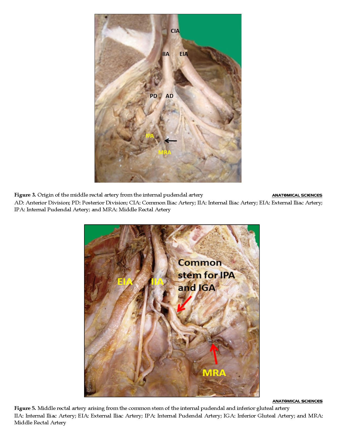
Parsons and Keith also studied the middle rectal artery and reported that the middle rectal artery might be present in the form of multiple vessels. When there is more than one vessel, they arise from the internal iliac artery, the inferior vesical artery, or the internal pudendal artery [5].
Hassen et al. explored the middle rectal artery. They found its origins from the posterior division of the internal iliac artery or internal pudendal artery [6].
The lack of a middle rectal artery was also reported in about 60% of cases by Lin M and associates [7]. In the present study, middle rectal artery existed in all cases.
Variable origins of the middle rectal artery given by Henry Hollinshead 2nd edition, volume 2 are shown in the following diagram. According to him, middle rectal artery may arise from internal pudendal artery, inferior gluteal artery, or obturator artery. It may also arise from the posterior part of internal iliac artery (Figure 6).
The middle rectal artery penetrates the fascia of rectum and laterally passes to the rectum postero. It is important in mesorectal excision in rectal carcinoma cases; as the middle rectal artery is usually removed in these cases. [8] Additionally, with advances in endoscopic surgery, the knowledge of precise anatomy of middle rectal artery is becoming more crucial for optimal rectal cancer surgery [9].
The middle rectal artery forms anastomoses with superior rectal artery. In low anterior resection of rectum, for rectal carcinoma, middle rectal artery is always removed. Thus, the knowledge of middle rectal artery and its variations is vital [10].
During the development of blood vessels, numerous primary capillary channels are formed. The most appropriate channels enlarge while the others are retracting or disappearing, which may result in the final arterial pattern. Unusual selection of channels from the primary capillaries might account for the anatomical variations affecting the arterial patterns [11].
During the development of blood vessels, numerous primary capillary channels are formed. The most appropriate channels enlarge while the others are retracting or disappearing, which may result in the final arterial pattern. Unusual selection of channels from the primary capillaries might account for the anatomical variations affecting the arterial patterns [11].
5. Conclusion
Variant origins of the middle rectal artery, like a branch from any of the branches of anterior division of internal iliac artery or the posterior division of internal iliac artery are commonly observed; thus, this artery is a surgically important vessel in rectal carcinoma excision procedures.
Ethical considerations
Compliance with ethical guidelines
All ethical principles were considered in this article.
Funding
This research did not receive any specific grant from funding agencies in the public, commercial, or not-for-profit sectors.
Authors' contributions
All authors contributed in preparing this article.
Conflict of interest
The authors declared no conflict of interest.
References
Gray H. The anatomical basis of clinical practice. 40th edition. London: Elsevier Churchill Livingstone; 2008.
Moore LM, Dally AF, Agur AMR. Clinically oriented anatomy. 6th edition. Philadelphia: Lippincott Williams & Wilkins; 2009.
Romanes GJ. Cunningham’s manual of practical anatomy. 15th edition. Oxford: Oxford Medical Publications; 1986.
Bergman RA, Thompson SA, Afifi AK, Saadeh FA. Compendium of human anatomic variation: Atlas and world literature. Baltimore and Munich: Urban & Schwarzenberg; 1988.
Parsons FG, Keith A. Sixth annual report of the Committee of Collective Investigation of the Anatomical Society of great Britain and Ireland. Mode of origin of the branches of the internal iliac artery. Journal of Anatomy and Physiology. 1897; 31:31-44.
Hentati H, Bouchiba N, Abderrazak, Mighri MM. Anatomy of superior and middle rectal arteries. 4th international symposium of Clinical and Applied Anatomy. 2012; 6:S1-S104. [DOI:10.2399/ana.12.001s]
Lin M, Chen W, Huang L, Ni J, Yin L. The anatomy of lateral ligament of rectum and its role in total mesorectal excision. World Journal Surgery. 2010; 34(3):594-8. [DOI:10.1007/s00268-009-0371-1] [PMID]
Kiyomatsu T, Ishihara S, Murono K, Otani K, Yasuda K, et al. Anatomy of the middle rectal artery: A review of historical literature. Surg Today. 2017; 47(1):14-9. [DOI: 10.1007/s00595-016-1359-8] [PMID]
LjA D, Diaz-Franco D, Schemainda R, Bezerra AJC. Morphology of the middle rectal arteries. Surgical and Radiological Anatomy. 1986; 8(4):229-36. [DOI:10.1007/BF02425072] [PMID]
Hollinshead WH. Anatomy for surgeons. 5th edition. New York: Harper & Row Publishers; 1978.
Adachi B, Hasebe K, Igakubu KD. Das arteriensystem der Japaner. Kyoto: Kaiserlich-japanische Universität zu Kyoto, in kommission bei “Maruzen Co”, Kyoto and Tokyo; 1928.
Type of Study: Original |
Subject:
Gross Anatomy
Received: 2019/02/7 | Accepted: 2019/08/16 | Published: 2020/01/1
Received: 2019/02/7 | Accepted: 2019/08/16 | Published: 2020/01/1
Send email to the article author
| Rights and permissions | |
 |
This work is licensed under a Creative Commons Attribution-NonCommercial 4.0 International License. |
Contact Information
Anatomical Sciences Journal (ASJ)
Negah Institute for Scientific Communication, No.15, Na'eemi St., Mirzaye Shirazi St., Tehran, Iran.
Publisher Tel : +9821 4535 5555;
+9821 4535 5000
Website: http://www.anatomyjournal.ir/
E-mail: anatomyjournal@gmail.com
