Sat, Apr 20, 2024
Volume 15, Issue 1 (Winter & Spring 2018)
ASJ 2018, 15(1): 25-30 |
Back to browse issues page

Download citation:
BibTeX | RIS | EndNote | Medlars | ProCite | Reference Manager | RefWorks
Send citation to:


BibTeX | RIS | EndNote | Medlars | ProCite | Reference Manager | RefWorks
Send citation to:
Goodarzi N, Akbari G, Razeghi Tehrani P. Zinc Chloride, A New Material for Embalming and Preservation of the Anatomical Specimens. ASJ 2018; 15 (1) :25-30
URL: http://anatomyjournal.ir/article-1-192-en.html
URL: http://anatomyjournal.ir/article-1-192-en.html


1- Department of Basic Sciences, Faculty of Veterinary Medicine, Razi University, Kermanshah, Iran.
2- Department of Basic Sciences, Faculty of Veterinary Medicine, University of Tabriz, Tabriz, Iran.
3- Department of Basic Sciences, Faculty of Veterinary Medicine, Karaj Branch, Islamic Azad University, Karaj, Iran.
2- Department of Basic Sciences, Faculty of Veterinary Medicine, University of Tabriz, Tabriz, Iran.
3- Department of Basic Sciences, Faculty of Veterinary Medicine, Karaj Branch, Islamic Azad University, Karaj, Iran.
Full-Text [PDF 444 kb]
(3409 Downloads)
| Abstract (HTML) (5507 Views)


Full-Text: (3534 Views)
1. Introduction
Cadavers and animal carcasses are used to teach macroscopic anatomy curriculum in medical and veterinary schools. One of the most important prerequisites for using human cadavers and animal carcasses is their proper preservation [1]. Thus, anatomical specimens must be protected from decomposition by some methods like embalming with certain chemical substances. Trying to embalm and preserve the human body dates back to 3000 years ago. Then, the main motive for embalming was religious beliefs. Many ancient cultures believed that eternal life was associated with an intact body and rotten bodies are deprived from the afterlife. According to this belief, cadavers do not corrupt if be preserved under certain conditions and using natural methods such as freezing and heat or cold drying [2].
Well-embalmed cadavers for teaching anatomy must have the following characteristics: the organs and tissues should be preserved for a long time with the minimal shrinkage, deformation, and hardening, as well as maintaining flexibility [3]. Formaldehyde is one of the most important chemicals commonly used for preservation of the cadavers. A German chemist, August Wilhelm von Hofmann, discovered formaldehyde in 1869 and Laskowski introduced phenol for embalming in the mid-19th century (1886) [4].
At that time, formaldehyde was recognized as an excellent preservative and used as a basic material for embalming solutions. The exact concentration of formaldehyde used was a controversial topic. Some recommended a concentration of 3% and some others 10%. In addition, side effects of formaldehyde such as skin and respiratory irritation, conjunctivitis, and headache was marked. So far, several modified formulae have been suggested for embalming in the literature. Kieserling method was introduced in 1897 for maintaining color and shapes and still is widely used. However, this method is mainly applicable for separated samples and is not suitable for anatomical dissection [5]. Jaurès presented another fixative solution containing chloral hydrate and formaldehyde [6]. In 1952, Woodburne and Lawrence applied a modified formulation with common compounds, including alcohol, glycerin, formaldehyde, and phenol [7].
Richins et al. [8] used a new solution for embalming that contains potassium pyrophosphate and magnesium chloride to reduce formaldehyde fixation-induced stiffness. Moreover, they replaced the phenol with sodium pentachlorophenate to eliminate unpleasant cadaver odor and look better its color. In 1983, Logan described a guidance for fixing the cadavers that was different from several aspects compared to the common methods. In this procedure, fresh cadaver is frozen at -35ºC, thawed for two days then the venous system is rinsed with a blood diluent and a preservative solution containing alcohol, glycerin, phenol, and finally a small amount of formaldehyde is injected in the arteries [9]. Thiel presented a new method for the preservation of the cadaver with natural color [10, 11].
The most important advantage of this method was satisfying the high standards of preservation without releasing harmful substances into the environment. However, this method is quite complex and includes several expensive materials. In this method, the flexibility of the preserved corpses is comparable with fresh ones and their color is also well preserved. These specimens can be used in various surgical trainings such as arthroscopy, laparoscopy, oral surgery, and implants [12, 13, 14]. Despite the diverse formulas that have been presented for preservation, formaldehyde is still used as the basic material. Considering the actual and potential negative effects of formaldehyde for the health of the students, teachers and staff who are exposed to it as well as environmental contamination, we attempted to examine a new solution containing high amounts of ZnCl2 salt for fixation and preservation of the anatomical specimens. The results of the present study would be helpful to introduce a cleaner and safer solution and consequently a better experience of dissection.
2. Materials and Methods
The embalming solution used in this study consisted of ZnCl2 powder, glycerin, and thymol. About 400 g of ZnCl2 salt was solved in 1 liter of water. Then, 1 liter of glycerin (emollient) and 200 g of thymol (anti-fungal) were added per 10 liters of the solution. An equine (dog), four carnivores (dog) and a small ruminant (goat) were used for embalming. Two dogs were embalmed with the solution described above and the two others were injected just with 40% ZnCl2 solution without using glycerin and thymol.
The animals were anesthetized by injecting ketamine and xylazine (5 mg/kg ketamine + 0.5 mg/kg xylazine IM in dogs, 5 mg/kg ketamine + 0.1 mg/kg xylazine IM in goat, 2 mg/kg ketamine + 1 mg/kg xylazine IV in dog). The carotid artery was identified through an almost 10-cm incision in the upper third of the neck and ligated with two scissors. Then a small incision was made on the arterial wall through which a cannula with the same diameter of the artery was inserted. Then, the lower scissor (which was near to the heart) was opened to allow blood to flow out through the cannula. After complete bleeding, 20, 5, and 5 liters of the solution was pumped into the carotid artery and the circulatory system of donkey, dogs, and goats, respectively. Pumping was stopped after viewing the foam in the body’s natural openings like the nostrils and anus and the artery was closed with a thread. At the end, the limbs extremities were covered by the cloths soaked in glycerin and the animals were transferred to the 4ºC refrigerator. Carnivorous and ruminant specimens were dissected after one week and equine specimen was dissected after three weeks.
3. Results
The results showed that 40% ZnCl2 solution can be properly used for embalming of animals. As in dissection, the muscular tissue and joints were found to be quite soft and flexible. Color of the muscles and internal organs was quite similar to those embalmed with com
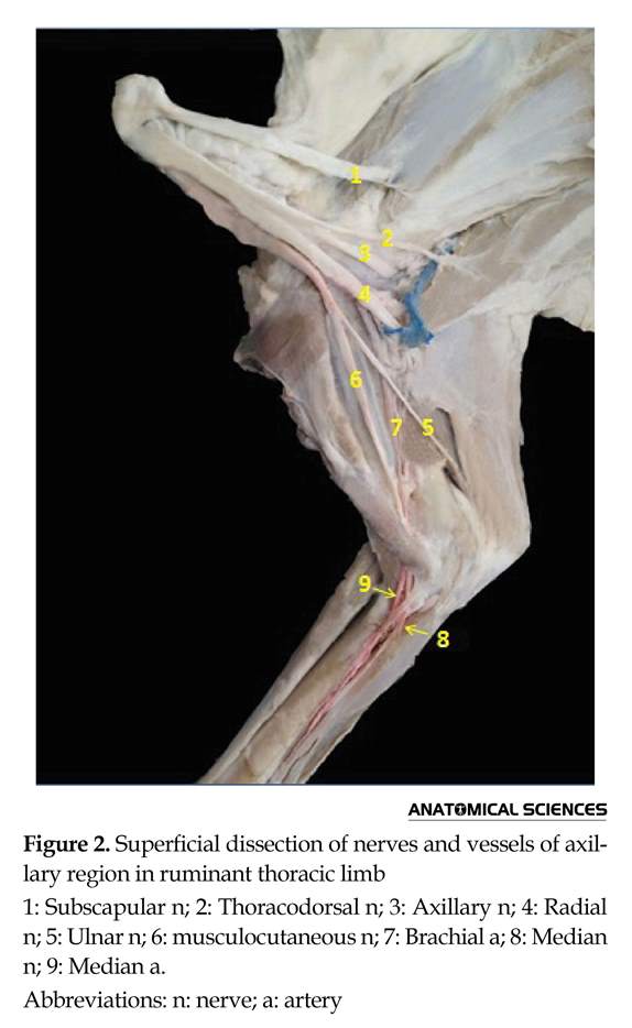
mon solution containing formalin (Figure 1). Vessels and nerves were easily dissected and detected (Figure 2, 3). There were no sign of drying and salt deposition over the skin or internal organs. It is worth noting that similar results were obtained in the cases which were preserved with 40% ZnCl2 solution without thymol and glycerin and there was no sign of fungal growth, too. Therefore, antifungal and softeners substances are not be needed.
4. Discussion
Concern is rising about the dangers of formaldehyde use in the workplace. On the other hand, the attempts to find a practical solution in accordance with the health and safety regulations is necessary for setting a standard limit for exposure to formaldehyde. The other complication is that university officials and health security of medical schools may be faced with major lawsuits if employees contact with excessive amounts of formaldehyde [15]. The chemical compounds that had been used for embalming for decades, now are inapplicable and impractical. The search for a solution with a small amount or without formaldehyde is an urgent issue. However, this
issue has been widely neglected and limited efforts have been made to reduce the concentration of formaldehyde or use a substitute in embalming solutions [16, 17]. In the present study, excellent results were obtained with complete elimination of formaldehyde and replace it with ZnCl2 salt.
The idea of using common salts as the main compound of the embalming solutions originates from its application in food industries. Salt has been used as a great and cheap preservative in food industries for centuries. High levels of salt in many processed foods such as meat, fish and vegetables are common and create an environment that minimizes the growth of bacteria and fungi. Ambroise Pare (1510-1590), an anatomist and one of the pioneers in surgical techniques, introduced the common salt as a component of his embalming solution [18]. Coleman and Cogan [3] offered a solution to embalm corpses. Their solution contains a high concentration of salt and small quantities of formaldehyde. Logan modified their solution with adding formaldehyde, glycerin, phenol, isopropyl alcohol and a large amount of sodium chloride. They stated that salt prevents the cadavers from excessive drying and maintains the natural color of tissues. Moreover, tissues remain soft and flexible while their deformation is minimized. The obtained results in the study confirm these findings, as muscles were quite soft and flexible and no sign of drying or salt decomposition was found in the specimens. However, the muscles color was gray similar to the color of formalin fixed samples. In addition, ordinary salt are available with low cost. Considering these features, it seems that saturated salts solution are very suitable for embalming animal corpses. Muscles make up 40%-50% of the body weight. Salted or cooked muscles remain soft and have a good resistance against drying and microbial contamination. In a study, various methods of embalming with saturated salt solution, Thiel solution, and traditional formaldehyde-based solution were compared and investigated for surgical training. Traditional methods based on formaldehyde, despite having benefits such as the least risk of infection and good quality for histological studies, have some disadvantages such as hardening of tissues and joints, change of tissues color, low quality of imaging and health risks (carcinogenic). Instead, by using a Thiel solution, tissues and joints remain soft and flexible with natural color and appearance and imaging capability is also high. However, this method is costly and very time consuming with difficult technique. On the other hand, the saturated salt solution maintains natural color of tissues, provides high quality images, and has low cost. The disadvantages of this solution for surgical training compared to Thiel solution are low flexibility and softness of tissues and joints and also edema formation, especially in the subcutaneous tissues [19].
The obtained results showed that 40% ZnCl2 solution can embalm and preserve anatomical specimens properly, i.e., the muscles and joints remain soft and flexible, besides vessels and nerves can be easily dissected and detected.
Acknowledgements
The authors are thankful to the Deputy of Research and Technology of Razi University for the financial support. Also, we highly appreciate Mrs. Dolati for her invaluable assistance in editing the manuscript.
Conflict of Interest
The authors declared no conflicts of interest.
References
- Brenner E, Maurer H, Moriggl B, Pomaroli A. The human cadaver as an educational tool – classification and comparison with other educational tools. Annals of Anatomy. 2003; 185: 229–30.
- Brenner E. Human body preservation - old and new techniques. Journal of Anatomy. 2014; 224(3):316–44. doi: 10.1111/joa.12160
- Coleman R, Kogan I. An improved low-formaldehyde embalming fluid to preserve cadavers for anatomy teaching. Journal of Anatomy. 1998; 192(3):443–6. doi: 10.1046/j.1469-7580.1998.19230443.x
- Ezugworie J, Anibeze C, Ozoemena R. Trends in the development of embalming methods. The Internet Journal of Alternative Medicine. 2009; 7(2). doi: 10.5580/29b
- Pulvertaft RJV. Museum techniques: A review. Journal of Clinical Pathology. 1950; 3(1):1–23. doi: 10.1136/jcp.3.1.1
- Bradbury SA, Hoshino K. An improved embalming procedure for long-lasting preservation of the cadaver for anatomical study. Cells Tissues Organs. 1978; 101(2):97–103. doi: 10.1159/000144954
- Woodburne RT, Lawrence CA. An improved embalming fluid formula. The Anatomical Record. 1952; 114(3):507–14. doi: 10.1002/ar.1091140309
- Richins CA, Roberts EC, Zeilmann JA. Improved fluids for anatomical embalming and storage. The Anatomical Record. 1963; 146(3):241–3. doi: 10.1002/ar.1091460309
- Logan B. The long-term preservation of whole human cadavers destined for anatomical study. Annals of The Royal College of Surgeons of England. 1983; 65(5): 333.
- Thiel W. [The preservation of whole bodies in natural colors (German)]. Annals of Anatomy - Anatomischer Anzeiger. 1992; 174(3):185–95. doi: 10.1016/s0940-9602(11)80346-8
- Thiel W. [Supplement for the preservation of whole bodies according to W (German)]. Annals of Anatomy - Anatomischer Anzeiger. 2002; 184(3):267–9. doi: 10.1016/s0940-9602(02)80121-2
- Groscurth P, Eggli P, Kapfhammer J, Rager G, Hornung J-P, Fasel JDH. Gross anatomy in the surgical curriculum in Switzerland: Improved cadaver preservation, anatomical models, and course development. The Anatomical Record. 2001; 265(6):254–6. doi: 10.1002/ar.10030
- Giger U, Frésard I, Häfliger A, Bergmann M, Krähenbühl L. Laparoscopic training on Thiel human cadavers: A model to teach advanced laparoscopic procedures. Surgical Endoscopy. 2007; 22(4):901–6. doi: 10.1007/s00464-007-9502-7
- Hölzle F, Franz E-P, Lehmbrock J, Weihe S, Teistra C, Deppe H, et al. Thiel embalming technique: A valuable method for teaching oral surgery and implantology. Clinical Implant Dentistry and Related Research. 2009; 14(1):121–6. doi: 10.1111/j.1708-8208.2009.00230.x
- Coleman R. Reducing the levels of formaldehyde exposure in gross anatomy laboratories. The Anatomical Record. 1995; 243(4):531–3. doi: 10.1002/ar.1092430417
- O'Sullivan E, Mitchell BS. An improved composition for embalming fluid to preserve cadavers for anatomy teaching in the United Kingdom. Journal of anatomy. 1993; 182(Pt 2):295-7. PMCID: PMC1259842
- Macdonald GJ, MacGregor DB. Procedures for embalming cadavers for the dissecting laboratory. Experimental Biology and Medicine. 1997; 215(4):363–5. doi: 10.3181/00379727-215-44144
- Mayer RG. Embalming: history, theory, and practice. New York: McGraw-Hill; 2012.
- Hayashi S, Naito M, Kawata S, Qu N, Hatayama N, Hirai S, et al. History and future of human cadaver preservation for surgical training: From formalin to saturated salt solution method. Anatomical Science International. 2015; 91(1):1–7. doi: 10.1007/s12565-015-0299-5
Type of Study: Original |
Subject:
Gross Anatomy
Received: 2016/05/6 | Accepted: 2017/10/3 | Published: 2018/01/1
Received: 2016/05/6 | Accepted: 2017/10/3 | Published: 2018/01/1
References
1. Brenner E, Maurer H, Moriggl B, Pomaroli A. The human cadaver as an educational tool – classification and comparison with other educational tools. Annals of Anatomy. 2003; 185: 229–30.
2. Brenner E. Human body preservation - old and new techniques. Journal of Anatomy. 2014; 224(3):316–44. doi: 10.1111/joa.12160 [DOI:10.1111/joa.12160]
3. Coleman R, Kogan I. An improved low-formaldehyde embalming fluid to preserve cadavers for anatomy teaching. Journal of Anatomy. 1998; 192(3):443–6. doi: 10.1046/j.1469-7580.1998.19230443.x [DOI:10.1046/j.1469-7580.1998.19230443.x]
4. Ezugworie J, Anibeze C, Ozoemena R. Trends in the development of embalming methods. The Internet Journal of Alternative Medicine. 2009; 7(2). doi: 10.5580/29b [DOI:10.5580/29b]
5. Pulvertaft RJV. Museum techniques: A review. Journal of Clinical Pathology. 1950; 3(1):1–23. doi: 10.1136/jcp.3.1.1 [DOI:10.1136/jcp.3.1.1]
6. Bradbury SA, Hoshino K. An improved embalming procedure for long-lasting preservation of the cadaver for anatomical study. Cells Tissues Organs. 1978; 101(2):97–103. doi: 10.1159/000144954 [DOI:10.1159/000144954]
7. Woodburne RT, Lawrence CA. An improved embalming fluid formula. The Anatomical Record. 1952; 114(3):507–14. doi: 10.1002/ar.1091140309 [DOI:10.1002/ar.1091140309]
8. Richins CA, Roberts EC, Zeilmann JA. Improved fluids for anatomical embalming and storage. The Anatomical Record. 1963; 146(3):241–3. doi: 10.1002/ar.1091460309 [DOI:10.1002/ar.1091460309]
9. Logan B. The long-term preservation of whole human cadavers destined for anatomical study. Annals of The Royal College of Surgeons of England. 1983; 65(5): 333.
10. Thiel W. [The preservation of whole bodies in natural colors (German)]. Annals of Anatomy - Anatomischer Anzeiger. 1992; 174(3):185–95. doi: 10.1016/s0940-9602(11)80346-8 [DOI:10.1016/S0940-9602(11)80346-8]
11. Thiel W. [Supplement for the preservation of whole bodies according to W (German)]. Annals of Anatomy - Anatomischer Anzeiger. 2002; 184(3):267–9. doi: 10.1016/s0940-9602(02)80121-2 [DOI:10.1016/S0940-9602(02)80121-2]
12. Groscurth P, Eggli P, Kapfhammer J, Rager G, Hornung J-P, Fasel JDH. Gross anatomy in the surgical curriculum in Switzerland: Improved cadaver preservation, anatomical models, and course development. The Anatomical Record. 2001; 265(6):254–6. doi: 10.1002/ar.10030 [DOI:10.1002/ar.10030]
13. Giger U, Frésard I, Häfliger A, Bergmann M, Krähenbühl L. Laparoscopic training on Thiel human cadavers: A model to teach advanced laparoscopic procedures. Surgical Endoscopy. 2007; 22(4):901–6. doi: 10.1007/s00464-007-9502-7 [DOI:10.1007/s00464-007-9502-7]
14. Hölzle F, Franz E-P, Lehmbrock J, Weihe S, Teistra C, Deppe H, et al. Thiel embalming technique: A valuable method for teaching oral surgery and implantology. Clinical Implant Dentistry and Related Research. 2009; 14(1):121–6. doi: 10.1111/j.1708-8208.2009.00230.x [DOI:10.1111/j.1708-8208.2009.00230.x]
15. Coleman R. Reducing the levels of formaldehyde exposure in gross anatomy laboratories. The Anatomical Record. 1995; 243(4):531–3. doi: 10.1002/ar.1092430417 [DOI:10.1002/ar.1092430417]
16. O'Sullivan E, Mitchell BS. An improved composition for embalming fluid to preserve cadavers for anatomy teaching in the United Kingdom. Journal of anatomy. 1993; 182(Pt 2):295-7. PMCID: PMC1259842 [PMID] [PMCID]
17. Macdonald GJ, MacGregor DB. Procedures for embalming cadavers for the dissecting laboratory. Experimental Biology and Medicine. 1997; 215(4):363–5. doi: 10.3181/00379727-215-44144 [DOI:10.3181/00379727-215-44144]
18. Mayer RG. Embalming: history, theory, and practice. New York: McGraw-Hill; 2012.
19. Hayashi S, Naito M, Kawata S, Qu N, Hatayama N, Hirai S, et al. History and future of human cadaver preservation for surgical training: From formalin to saturated salt solution method. Anatomical Science International. 2015; 91(1):1–7. doi: 10.1007/s12565-015-0299-5 [DOI:10.1007/s12565-015-0299-5]
Send email to the article author
| Rights and permissions | |
 |
This work is licensed under a Creative Commons Attribution-NonCommercial 4.0 International License. |
Contact Information
Anatomical Sciences Journal (ASJ)
Negah Institute for Scientific Communication, No.15, Na'eemi St., Mirzaye Shirazi St., Tehran, Iran.
Publisher Tel : +9821 4535 5555;
+9821 4535 5000
Website: http://www.anatomyjournal.ir/
E-mail: anatomyjournal@gmail.com
